
Series
|

|
|
|
Preparation |
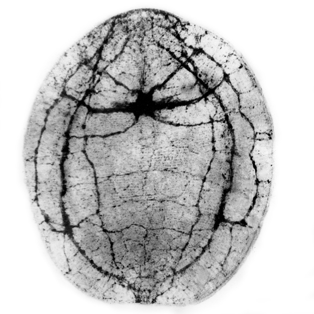
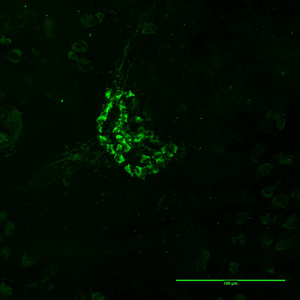
Description: FMRFamid immunoreactivity in sensory tentacles of Polypodium hydriforme (Cnidaria, Polypodiozoa). Organs: nerve plexus, neuron(s), musculature innervation / musculature Methods: Immunohistochemistry - FMRFamide |
|
Series
|

|
|
|
Preparation |
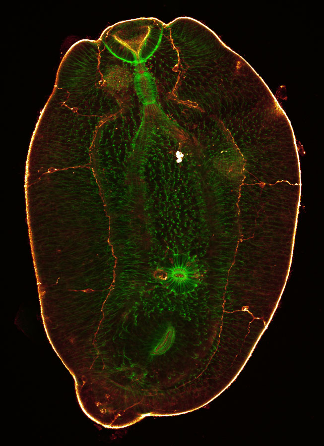
Description: Confocal scanning laser microscope image of double-stained specimens. GYIRFamide pattern is shown in red; 5-HT pattern is shown in green. Ventral side, optical sections taken from the level of the ventral epidermis to the level of the bilithophorous statocyst, including the latter. Note the extensive development of GYIRFamide-IR fibres on the ventral side and the presence of large neurons associated with the ventral part of the ring. Organs: сentral nervous system, nerve cord, nerve plexus, commissure, nerve, neuron(s) Methods: Immunohistochemistry - serotonin, |
|
Series
|

|
|
|
Preparation |
Description: Dorsal view. One can see the brain and 3 pairs of longitudinal nerve cords (dorsal, dorsolateral and lateral). Thicker proximal parts of the longitudinal cords form the commissural brain. Fibers of submuscular nerve plexus are evenly distributed over the body. Organs: сentral nervous system, nerve cord, nerve plexus, cerebral brain or its part, peripheral nervous system Methods: Histochemistry - revealing of choline esterases after Gerebtzoff |
|
Series
|

|
|
|
Preparation |
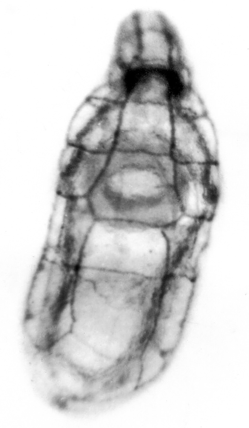
Description: Confocal scanning laser images of phalloidin stained (in red) specimen with FMRFamide (green) immunoreactive labeling. FMRF-IR is visible in the brain neuropile (n) and in the sensory cells of the sensory pit (short arrow). Phalloidin stains thick longitudinal muscles and thin transverse muscles (long arrow) between the frontal lobes of the brain. Organs: сentral nervous system, cerebral brain or its part, neuron(s), receptor cell(s), musculature innervation / musculature, Sensory organ Methods: Immunohistochemistry - FMRFamide, |
|
Series
|

|
|
|
Preparation |
Description: Ventral view. Image shows the brain and 3 pairs of longitudinal nerve cords (dorsal, ventral and lateral). Note many circular commissures between the cords. Fibers of submuscular nerve plexus are evenly distributed over the body. Organs: сentral nervous system, nerve cord, nerve plexus, cerebral brain or its part, commissure, nerve, neural tract, peripheral nervous system Methods: Histochemistry - revealing of choline esterases after Gerebtzoff |
|
Series
|

|
|
|
Preparation |
Description: Dorsal view. Image shows one pair of longitudinal dorsal nerve cords that produce numerous branches. Fibers of submuscular nerve plexus are evenly distributed over the body. Organs: nerve cord, nerve plexus, peripheral nervous system Methods: Histochemistry - revealing of choline esterases after Gerebtzoff |
|
Series
|

|
|
|
Preparation |
Description: Ventral view. Image shows one pair of longitudinal ventral nerve cords that produce numerous branches. Fibers of submuscular nerve plexus are evenly distributed over the body. Organs: nerve cord, nerve plexus, peripheral nervous system Methods: Histochemistry - revealing of choline esterases after Gerebtzoff |
|
Series
|

|
|
|
Preparation |
Description: Catecholaminergic neurons of the brain are distributed around the eyes. The image also shows the neuropile of the brain. Organs: сentral nervous system, cerebral brain or its part, neuron(s) Methods: Histochemistry - catecholaminergic nervous elements (GIF) |
|
Series
|
|
|
|
Preparation |
Description: Ventral view. Image shows the brain, frontal nerve trunk and three pairs of longitudinal nerve cords, six circular commissures. Fibers of submuscular nerve plexus are evenly distributed over the body. Organs: сentral nervous system, nerve cord, nerve plexus, cerebral brain or its part, commissure, peripheral nervous system Methods: Histochemistry - revealing of choline esterases after Gerebtzoff |
|
Series
|

|
|
|
Preparation |
Description: Dorsal view. Image shows the brain, two pairs of longitudinal nerve cords, the elliptical structure in the neuropile of the brain, brain neurons. Organs: сentral nervous system, nerve cord, cerebral brain or its part, neuron(s) Methods: Immunohistochemistry - FMRFamide, |
|
Series
|

|
|
|
Preparation |
Description: Ventral view. Image shows the brain, two pairs of longitudinal nerve cords, brain neurons. Organs: сentral nervous system, nerve cord, cerebral brain or its part, neuron(s) Methods: Immunohistochemistry - FMRFamide, |
|
Series
|

|
|
|
Preparation |
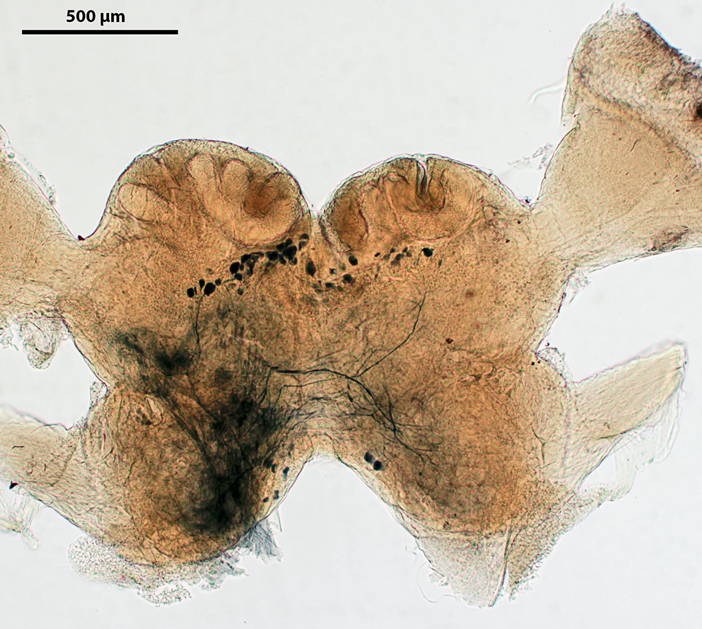
Description: General view of a 22-day-old metacercaria of Diplostomum pseudospathaceum (Platyhelminthes) showing the serotonergic nervous system and the musculature. The series shows the brain (cerebral ganglia), the anterior nerves and longitudinal nerve cords that extend from the brain, the commissures connecting the nerve cords and the nerves innervating the internal organs (suckers, lappets and the holdfast). Organs: сentral nervous system, nerve cord, cerebral brain or its part, ganglion / ganglia, commissure, nerve, neuron(s), larval nervous system, musculature innervation / musculature, peripheral nervous system Methods: Immunohistochemistry - serotonin, |
|
Series
|

|
|
|
Preparation |
Description: General view of a 22-day-old metacercaria of Diplostomum pseudospathaceum (Platyhelminthes) showing the serotonergic nervous system and the musculature. The series shows the brain (cerebral ganglia), the anterior nerves and longitudinal nerve cords that extend from the brain, the commissures connecting the nerve cords and the nerves innervating the internal organs (sucker, lappets and the holdfast). Organs: сentral nervous system, nerve cord, cerebral brain or its part, ganglion / ganglia, commissure, nerve, larval nervous system, musculature innervation / musculature, peripheral nervous system Methods: Immunohistochemistry - FMRFamide, |
|
Series
|
|
|
|
Preparation |
Description: General view of a 22-day-old metacercaria of Diplostomum pseudospathaceum (Trematoda, Platyhelminthes) showing the musculature and substance P-immunoreactive nervous elements. The series shows nervous elements in the cerebral ganglia, in the anterior nerves innervating the oral sucker, in the ventral longitudinal nerve cords and in the neurons innervating the tegument and hindbody. Organs: сentral nervous system, nerve cord, cerebral brain or its part, nerve, neuron(s), larval nervous system, musculature innervation / musculature, peripheral nervous system Methods: Immunohistochemistry - substance P, |
|
Series
|

|
|
|
Preparation |
Description: Dorsal view. Image shows the brain, longitudinal dorsal nerve cords, circular commissures. Organs: сentral nervous system, nerve cord, cerebral brain or its part, ganglion / ganglia, commissure, neural tract Methods: Histochemistry - revealing of choline esterases after Gerebtzoff |
|
Series
|
|
|
|
Preparation |
Description: Ventral view. Image shows the brain, longitudinal ventral nerve cords, circular commissures. Organs: сentral nervous system, nerve cord, cerebral brain or its part, ganglion / ganglia, commissure, neural tract Methods: Histochemistry - revealing of choline esterases after Gerebtzoff |
|
Series
|

|
|
|
Preparation |
Description: Ventral view. Image shows the brain, longitudinal ventral and dorsal nerve cords, circular commissures, innervation of oral and ventral suckers. Organs: сentral nervous system, nerve cord, cerebral brain or its part, commissure, neural tract Methods: Histochemistry - revealing of choline esterases after Gerebtzoff |
|
Series
|

|
|
|
Preparation |
Description: Ventral view. Image shows ganglia, longitudinal ventral nerve cords, and submuscular nerve plexus. Organs: сentral nervous system, nerve cord, nerve plexus, ganglion / ganglia, peripheral nervous system Methods: Histochemistry - revealing of choline esterases after Gerebtzoff |
|
Series
|

|
|
|
Preparation |
Description: Ventral view. Image shows the brain, longitudinal nerve cords, circular commissures, innervation of the oral sucker. Organs: сentral nervous system, nerve cord, nerve plexus, cerebral brain or its part, commissure Methods: Histochemistry - revealing of choline esterases after Gerebtzoff |
|
Series
|

|
|
|
Preparation |
Description: Image shows the cholinergic nerve plexus in a bothrium. Organs: nerve plexus Methods: Histochemistry - revealing of choline esterases after Gerebtzoff |
|
Series
|

|
|
|
Preparation |
Description: Image shows the ring of cholinergic fibers in the scolex, and 4 bothria around the scolex. Organs: сentral nervous system, nerve cord, nerve plexus, cerebral brain or its part Methods: Histochemistry - revealing of choline esterases after Gerebtzoff |
|
Series
|

|
|
|
Preparation |
Description: Ventral view. Image shows longitudinal nerve cords, circular commissures, submuscular nerve plexus. Organs: nerve cord, nerve plexus, commissure, peripheral nervous system Methods: Histochemistry - revealing of choline esterases after Gerebtzoff |
|
Series
|

|
|
|
Preparation |
Description: Image shows longitudinal nerve cords, circular commissures, inner, submuscular and superficial nerve plexuses. Organs: сentral nervous system, nerve cord, nerve plexus, commissure Methods: Histochemistry - revealing of choline esterases after Gerebtzoff |
|
Series
|

|
|
|
Preparation |
Description: Image shows longitudinal nerve cords, circular commissures, inner and submuscular nerve plexuses. Organs: сentral nervous system, nerve cord, nerve plexus, commissure Methods: Histochemistry - revealing of choline esterases after Gerebtzoff |
|
Series
|

|
|
|
Preparation |
Description: Image shows longitudinal nerve cords, inner and submuscular nerve plexuses. Organs: сentral nervous system, nerve cord, nerve plexus Methods: Histochemistry - revealing of choline esterases after Gerebtzoff |
|
Series
|

|
|
|
Preparation |
Description: Image shows longitudinal nerve cords, submuscular nerve plexus. Organs: сentral nervous system, nerve cord, nerve plexus, larval nervous system Methods: Histochemistry - revealing of choline esterases after Gerebtzoff |
|
Series
|

|
|
|
Preparation |
Description: Image shows longitudinal nerve cords, circular comissures, submuscular nerve plexus. Organs: сentral nervous system, nerve cord, nerve plexus, commissure Methods: Histochemistry - revealing of choline esterases after Gerebtzoff |
|
Series
|

|
|
|
Preparation |
Description: Image shows longitudinal nerve cords, circular comissures, submuscular nerve plexus. Organs: сentral nervous system, nerve cord, nerve plexus, cerebral brain or its part, peripheral nervous system Methods: Histochemistry - revealing of choline esterases after Gerebtzoff |
|
Series
|

|
|
|
Preparation |
Description: Image shows longitudinal nerve cords, submuscular nerve plexus. Organs: сentral nervous system, nerve cord, nerve plexus, cerebral brain or its part, peripheral nervous system Methods: Histochemistry - revealing of choline esterases after Gerebtzoff |
|
Series
|

|
|
|
Preparation |
Description: The anterior body end of the turbellarian Microdalyellia picta (Rhabditophora, Platyhelminthes) showing the musculature of the body wall and pharynx and the nervous system stained with antibodies to alpha-tubulin. The series shows the brain, three pairs of longitudinal nerve cords (ventral, lateral and dorsal) that arise from the brain and the pharyngeal nervous system (nerve ring of the juncture line, longitudinal nerves that extend from the ring in the anterior and posterior directions, transverse and circular nerves of the pharynx and neurites running to the receptor cells of the pharyngeal papillae). Organs: сentral nervous system, nerve cord, nerve plexus, cerebral brain or its part, commissure, musculature innervation / musculature, nerve, receptor cell(s), visceral nervous system Methods: Immunohistochemistry - alpha-tubulin, |
|
Series
|

|
|
|
Preparation |
Description: Anterior body end of the turbellarian Monocelis fusca (Rhabditophora, Platyhelminthes) showing the serotonergic nervous system, the body-wall musculature and the muscular sheath of the brain. The series shows the brain, two pairs of longitudinal nerve cords (one lateral and one ventrolateral) that extend posteriorly from the brain, the anterior nerves (one pair of lateral nerves and one unpaired medial nerve) and the subepidermal nerve plexus. Organs: сentral nervous system, nerve plexus, cerebral brain or its part, ganglion / ganglia, nerve, neuron(s), musculature innervation / musculature, peripheral nervous system Methods: Immunohistochemistry - serotonin, |
|
Series
|

|
|
|
Preparation |
Description: Serotonergic nervous system and the musculature of the turbellarian Monocelis fusca (Rhabditophora, Platyhelminthes) in the region of the brain showing serotonergic elements in the brain neuropil, cell bodies of brain neurons, two pairs of londitudinal nerve cords (one lateral and one ventro-lateral) that extend forward from the brain and the nervous elements of the subepidermal plexus. The series also shows a pair of neurites extending from the brain to the statocyst (which is visible as a dark spot at the anterior end of the brain). Also visible are the body-wall musculature, the muscle sheath around the brain and the muscle fibers passing through the brain. Organs: сentral nervous system, cerebral brain or its part, nerve, neuron(s), musculature innervation / musculature Methods: Immunohistochemistry - serotonin, |
|
Series
|

|
|
|
Preparation |
Description: Platyias patulus, patterns of FMRFamide (red) and of 5-HT (green) immunoreactivity. General FMRFamide-IR pattern and 5-HT-IR pattern in a double-stained specimen are annotated separately. Note the ventrolateral nerve cords and anterior dorsal semi-ring (adr) with neurons (n). Organs: сentral nervous system, cerebral brain or its part, neural tract, connective, commissure, neuron(s) Methods: Immunohistochemistry - FMRFamide, |
|
Series
|

|
|
|
Preparation |
Description: Distribution of FMRFamide and serotonin- (5HT) immunoreactive elements in the brain and one of the longitudinal nerve cords. This series was the first that showed the distribution of these elements in the nervous system of rotifers. Organs: сentral nervous system, nerve cord, cerebral brain or its part, nerve, neuron(s) Methods: Immunohistochemistry - FMRFamide, |
|
Series
|

|
|
|
Preparation |
Description: Distribution of FMRFamide-immunoreactive elements in the brain and in one of the longitudinal nerve cords. Organs: сentral nervous system, nerve cord, cerebral brain or its part, neuron(s) Methods: Immunohistochemistry - FMRFamide |
|
Series
|

|
|
|
Preparation |
Description: Distribution of serotonin-immunoreactive elements in the brain and in one of the longitudinal nerve cords. Organs: сentral nervous system, nerve cord, cerebral brain or its part, nerve, neuron(s) Methods: Immunohistochemistry - serotonin |
|
Series
|

|
|
|
Preparation |
Description: 5HT-IR elements in the central nervous system of an embryo, iside the mother's body. You can see the neuropile, brain neurons, ventro-lateral cord. Organs: сentral nervous system, cerebral brain or its part, neuron(s), nerve cord Methods: Immunohistochemistry - FMRFamide |
|
Series
|

|
|
|
Preparation |
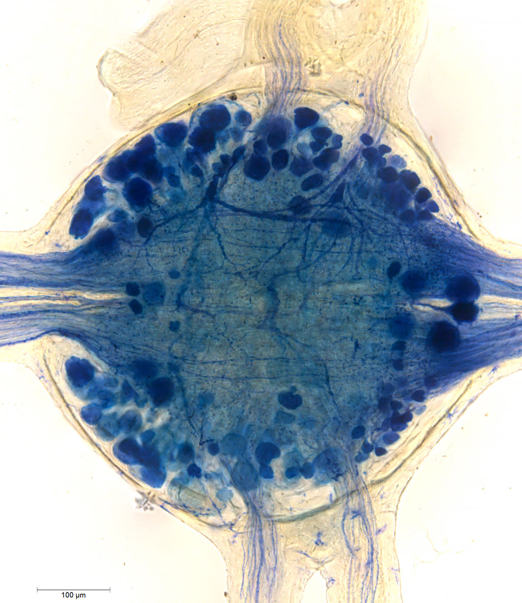
Description: Cristatella mucedo. Bottom view of the anal portion of the lophophore. Central nervous system of a zooid stained with antibodies to serotonin. The series shows the shape of the serotonin-positive part of the cerebral ganglion and large nerve cords innervating the horns of the lophophore. Organs: сentral nervous system, cerebral brain or its part, ganglion / ganglia, neural tract, peripheral nervous system, musculature innervation / musculature Methods: Immunohistochemistry - serotonin, |
|
Series
|

|
|
|
Preparation |
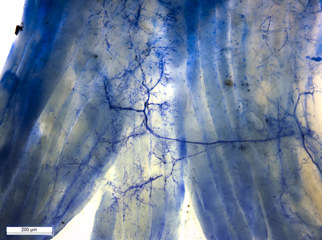
Description: Plumatella repens. Visceral and peripheral nervous systems at the base of the lophophore and in the wall of the cystide stained with antibodies to FMRFamide. The series shows that the majority of nerve fibers run within the intestinal wall and the wall of the cystide parallel to the apico-basal axis of the zooid. The preparations stained with phalloidin show the differences in muscle arrangement in different regions of the zooid and in orientation of muscle fibers in the walls of the cystide and intestine. Organs: neural tract, peripheral nervous system, visceral nervous system Methods: Histochemistry - F-actin in muscle fibers (Phalloidin), |
|
Series
|

|
|
|
Preparation |
Description: Fredericella sultana. Central nervous system and the nervous system of the lophophore stained with antibodies to serotonin. The lophophore of the zooid was imaged from the anal side. Deep in the preparation in the region of the cerebral ganglia, there is a clearly visible nerve net of serotonergic fibers. An additional staining with antibodies to alpha-tubulin shows the arrangement of nerves that innervate the base of the lophophore and the tentacles of the zooid. Phalloidin staining shows the positional relationships between the muscle and nerve fibers in the lophophore. Organs: neural tract, ganglion / ganglia, neuron(s), peripheral nervous system, musculature innervation / musculature, receptor cell(s) Methods: Histochemistry - F-actin in muscle fibers (Phalloidin), |
|
Series
|
|
|
|
Preparation |
Description: Zooid of a bryozoan with the central nervous system stained with antibodies to serotonin showing the shape of the serotonin-immunoreactive part of the cerberal ganglion and large nerve cords innervating the horns of the lophophore. Organs: сentral nervous system, ganglion / ganglia, nerve cord, nerve Methods: Immunohistochemistry - serotonin, |
|
Series
|
|
|
|
Preparation |
Description: Cerebral ganglion stained with antibodies to FMRFamide. The FMRFamide-immunoreactive part of the ganglion is represented by neuronal somata in the basal portion of the ganglion grouped as two symmetric rings with a commissure in the middle. Organs: сentral nervous system, ganglion / ganglia, commissure, neuron(s) Methods: Immunohistochemistry - FMRFamide, |
|
Series
|

|
|
|
Preparation |
Description: Preparation of a polypide of Plumatella repens stained with phalloidin and with antibodies to FMRFamide and tubulin showing multipolar neurons located in the nerve plexus of the body wall and the transverse fibers of the musculature. The lophophore is inside the polypide. Organs: peripheral nervous system, neuron(s), receptor cell(s), musculature innervation / musculature Methods: Immunohistochemistry - FMRFamide, |
|
Series
|

|
|
|
Preparation |
Description: Preparation of the lophophore base of Fredericella sultana stained with antibodies to alpha-tubulin. Organs: сentral nervous system, nerve cord, neural tract, nerve Methods: Immunohistochemistry - alpha-tubulin |
|
Series
|

|
|
|
Preparation |
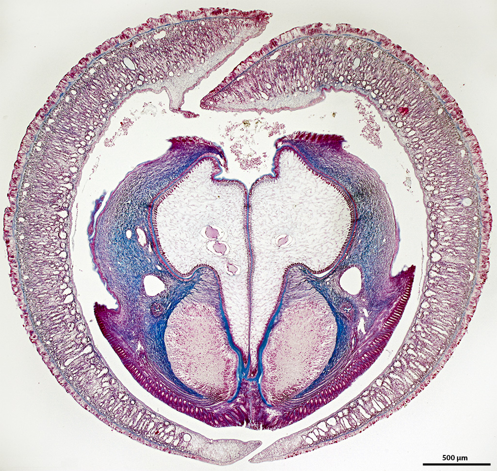
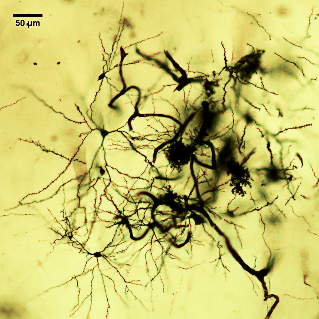
Description: A transverse section through four cerebral ganglia and lateral cords, which pass into the cerebral sensory organA transverse section through four cerebral ganglia and lateral cords, which pass into the cerebral sensory A transverse section through four cerebral ganglia and lateral cords, which pass into the cerebral sensory organ. Organs: сentral nervous system, ganglion / ganglia, neuron(s), commissure Methods: Neurohistology - Golgi-Colonnier method of silver impregnation of nervous elements |
|
Series
|

|
|
|
Preparation |
Description: Distribution of catecholaminergic nerve cells and fibers in the lateral nerve cords, cerebral ganglia, their commissures and connectives, and in the nerves that innervate the head and the digestive tract. Organs: сentral nervous system, nerve cord, cerebral brain or its part, ganglion / ganglia, connective, commissure, neural tract, nerve, neuron(s) Methods: Histochemistry - catecholaminergic nervous elements (GIF) |
|
Series
|

|
|
|
Preparation |
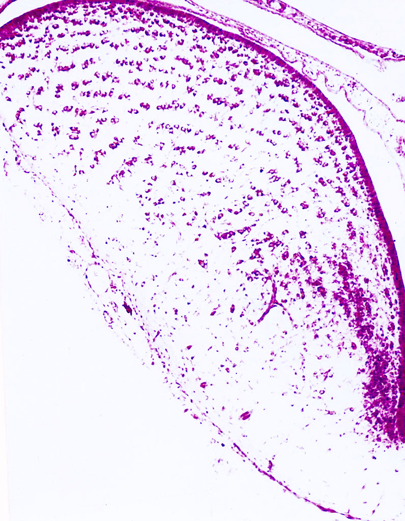
Description: Portion of the procerebrum (olfactory center) of a Roman snail. The series shows the region of the somata of the globuli cells (the main cellular elements of the procerebrum), the inner neuropil and the root of the tentacular nerve. Some of the globuli cells are grouped in columnar structures. Organs: сentral nervous system, cerebral brain or its part, ganglion / ganglia, neuron(s) Methods: Neurohistology - Golgi-Colonnier method of silver impregnation of nervous elements |
|
Series
|

|
|
|
Preparation |
Description: Synaptic contact between an incoming nerve fiber and the soma of a globular cell (axo-somatic synapse) in the region of cell bodies of the procerebrum. Organs: сentral nervous system, cerebral brain or its part, ganglion / ganglia, neuron(s) Methods: Electron microscopy - SEM |
|
Series
|

|
|
|
Preparation |
Description: Optical sections through the CNS and the ventral part of the animal showing the cerebral, pleural, pedal, and buccal ganglia, the 1st to 3rd pedal nerves and the innervation and the nerve cells of the anterior and middle portions of the foot. Organs: сentral nervous system, cerebral brain or its part, ganglion / ganglia, connective, commissure, nerve, neuron(s), receptor cell(s), peripheral nervous system Methods: Histochemistry - catecholaminergic nervous elements (GIF) |
|
Series
|
|
|
|
Preparation |
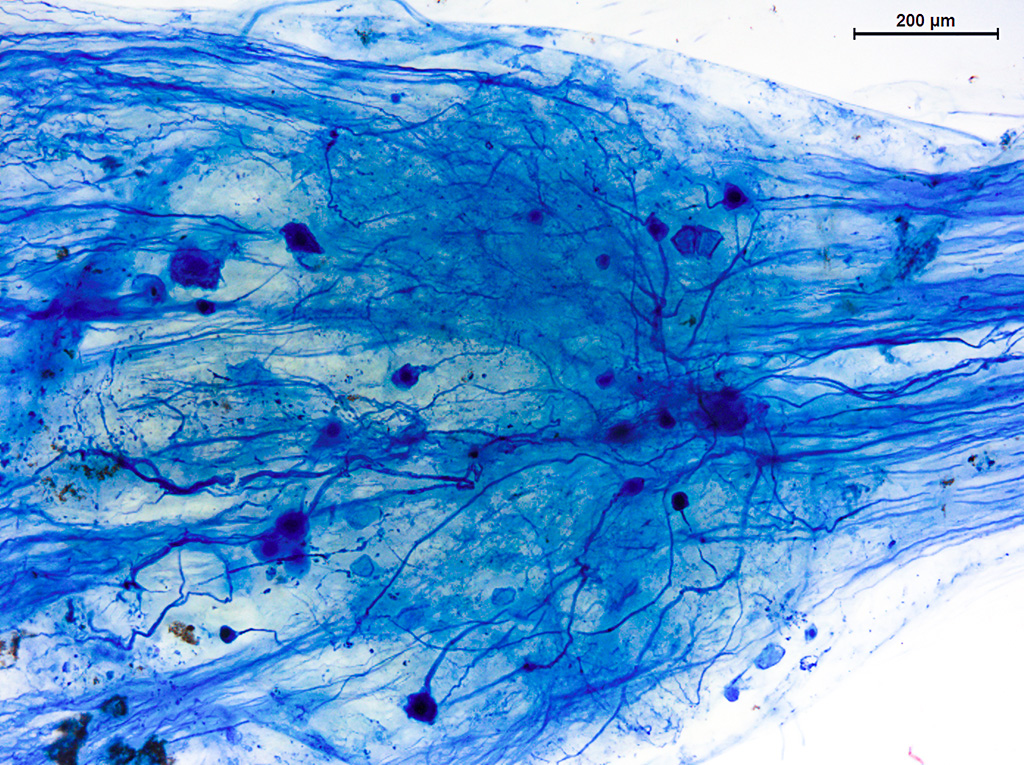
Description: Elements of the motor neuropil in the ganglion of the ventral nerve cords of a leech. The series shows a dendritic arborization typical for the motor neurons of leeches, which is located ipsilaterally in the neuropil of the ganglion. The series also shows fibers of the sensory neuropil passing through the ganglion inside the connectives. Organs: сentral nervous system, nerve cord, dorsal (spinal) cord or ventral cord, ganglion / ganglia, connective, nerve, neuron(s) Methods: Neurohistology - supravital methylene blue staining |
|
Series
|
|
|
|
Preparation |
Description: Branching (including terminal) of a motoneuron process on muscular fibers and neuromuscular contacts are seen. Organs: neuron(s) Methods: Neurohistology - supravital methylene blue staining |
|
Series
|

|
|
|
Preparation |
Description: Series of images of a body region of the polychete Protodrilus flavocapitatus (Polychaeta, Annelida) showing the muscles of the body wall and setae and the nervous system stained with antibodies to acetylated alpha-tubulin and FMRFamide. The series clearly shows two ventral nerve cords, the trasverse and diagonal commissures interconnecting the cords and the lateral nerves extending from the cords. The images also show cilia in the protonephridial canals. Organs: сentral nervous system, nerve cord, ganglion / ganglia, connective, commissure, nerve, neuron(s), musculature innervation / musculature, peripheral nervous system Methods: Immunohistochemistry - acetylated alpha-tubulin, |
|
Series
|
|
|
|
Preparation |
Description: Cross section at the level of the border between the obturacular and vestimental regions of the body. Organs: cerebral brain or its part Methods: Standard histology - Heidenhain's azan |
|
Series
|

|
|
|
Preparation |
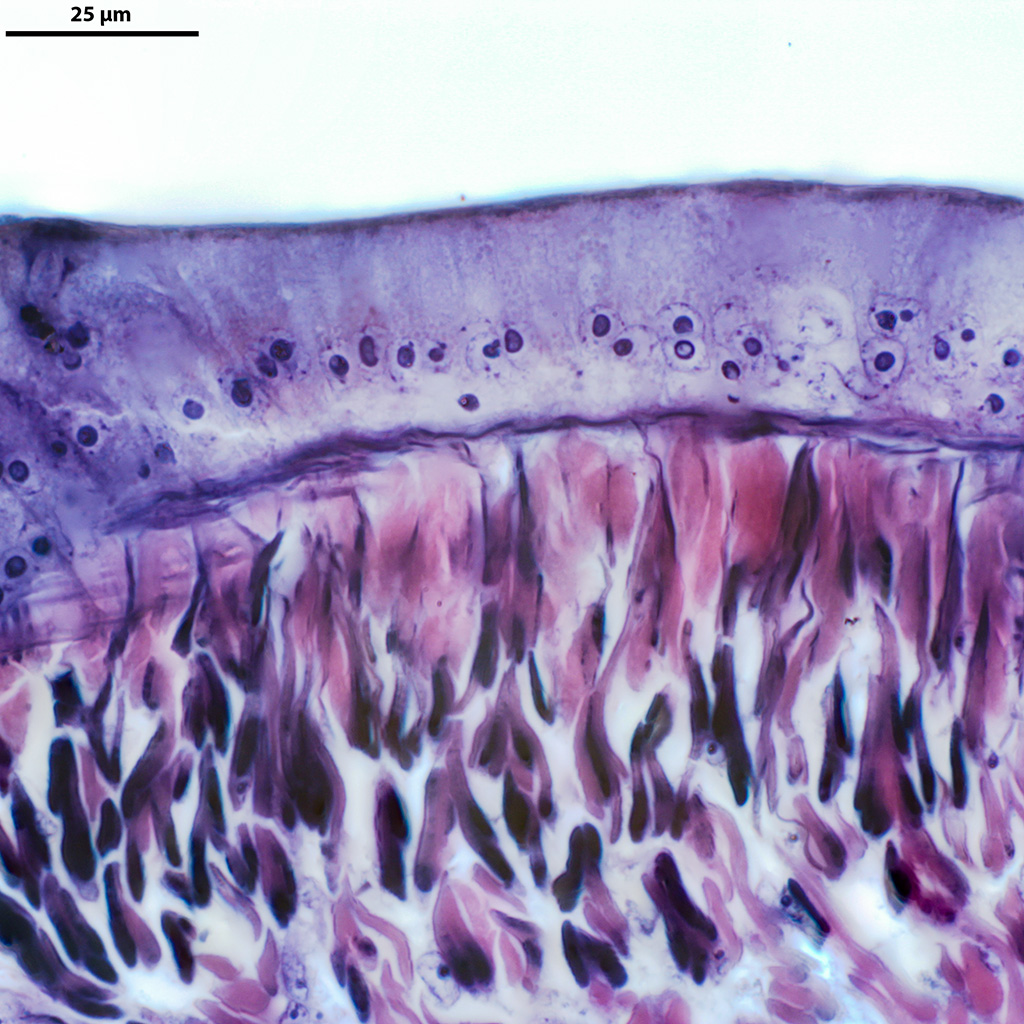
Description: Cross section of the tentacular crown of Spirobrachia grandis, stained with iron haematoxylin and eosin. Organs: nerve, receptor cell(s) Methods: Standard histology - hematoxylin, |
|
Series
|

|
|
|
Preparation |
Description: Cross section of the tentacular crown of Spirobrachia grandis, stained with iron haematoxylin and eosin. Organs: nerve, receptor cell(s) Methods: Standard histology - hematoxylin, |
|
Series
|

|
|
|
Preparation |
Description: Сross section of a tentacle in the tentacular crown of Spirobrachia grandis. Organs: nerve, receptor cell(s) Methods: Standard histology - hematoxylin |
|
Series
|

|
|
|
Preparation |
Description: Cross section of the tentacular crown of Spirobrachia grandis, stained with iron haematoxylin. Organs: nerve, receptor cell(s) Methods: Standard histology - hematoxylin |
|
Series
|

|
|
|
Preparation |
Description: Cross section through the tentacular crown of Polybrachia annulata. Organs: nerve, receptor cell(s) Methods: Standard histology - Mallory staining |
|
Series
|

|
|
|
Preparation |
Description: Cross section through the tentacular crown of Polybrachia annulata. Organs: nerve, receptor cell(s) Methods: Standard histology - Mallory staining |
|
Series
|

|
|
|
Preparation |
Description: Cross section through the tentacular crown of Polybrachia annulata. Organs: nerve, receptor cell(s) Methods: Standard histology - Mallory staining |
|
Series
|

|
|
|
Preparation |
Description: Oblique transverse section of the middle part of the trunk of Lamellisabella zachsi, stained with iron haematoxylin. Organs: nerve cord Methods: Standard histology - hematoxylin, |
|
Series
|

|
|
|
Preparation |
Description: Oblique transverse section of the middle part of the trunk of Lamellisabella zachsi, stained with iron haematoxylin. Organs: nerve cord Methods: Standard histology - hematoxylin, |
|
Series
|

|
|
|
Preparation |
Description: Oblique transverse section of the middle part of the trunk of Lamellisabella zachsi, stained with iron haematoxylin. Organs: nerve cord Methods: Standard histology - hematoxylin, |
|
Series
|

|
|
|
Preparation |
Description: Сross section of the tentacular crown of Spirobrachia grandis. Organs: nerve, receptor cell(s) Methods: Standard histology - Heidenhain's azan |
|
Series
|

|
|
|
Preparation |
Description: Oblique transverse section through the middle part of the trunk of Lamellisabella zachsi. Organs: nerve cord Methods: Standard histology - hematoxylin, |
|
Series
|
|
|
|
Preparation |
Description: Oblique transverse section through the middle part of the trunk of Lamellisabella zachsi. Organs: nerve cord Methods: Standard histology - hematoxylin, |
|
Series
|

|
|
|
Preparation |
Description: Oblique transverse section through the middle part of the trunk of Lamellisabella zachsi. Organs: nerve cord Methods: Standard histology - hematoxylin, |
|
Series
|

|
|
|
Preparation |
Description: none Organs: Sensory organ Methods: Standard histology - сarmine |
|
Series
|

|
|
|
Preparation |
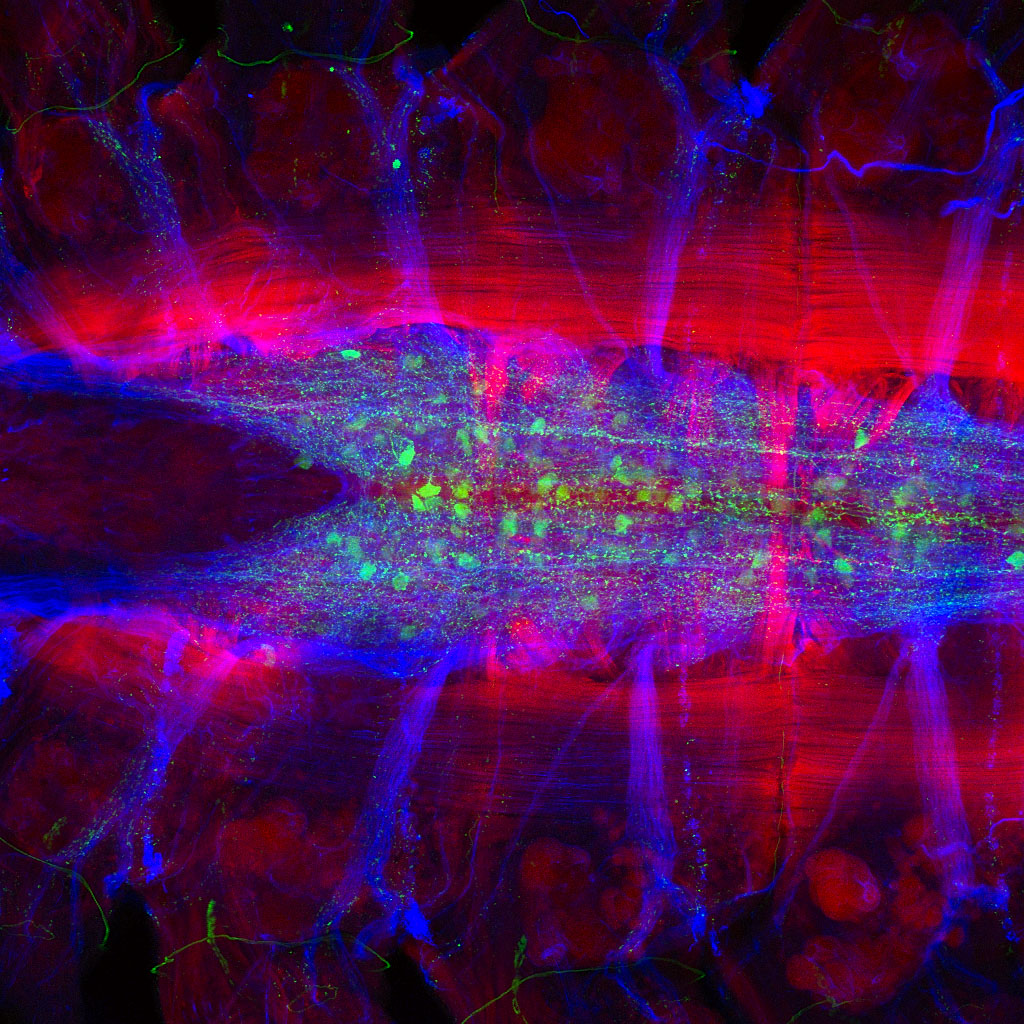
Description: Muscles are shown in red, ciliary structure and nerve fibers immunoreactive to acetylated tubulin are green. Autofluorescent chaete and serotonin-immunoreactive elements are cyan. Organs: сentral nervous system, cerebral brain or its part, ganglion / ganglia, nerve, peripheral nervous system Methods: Immunohistochemistry - serotonin, |
|
Series
|

|
|
|
Preparation |
Description: Muscles are shown in red, FMRFamide-positive immunoreactivity is green. Nerve fibers, labelled by acetylated tubulin, are shown in blue. Organs: сentral nervous system, cerebral brain or its part, ganglion / ganglia, Sensory organ Methods: Immunohistochemistry - FMRFamide, |
|
Series
|

|
|
|
Preparation |
Description: Muscles are shown in red, serotonin-positive immunoreactivity is green. Nerve fibers, labelled by acetylated tubulin, are shown in magenta. Organs: сentral nervous system, ganglion / ganglia, connective, nerve Methods: Immunohistochemistry - serotonin, |
|
Series
|
|
|
|
Preparation |
Description: Muscles are shown in red, serotonin-positive immunoreactivity is green. Nerve fibers, labelled by acetylated tubulin, are shown in blue. Organs: сentral nervous system, ganglion / ganglia, connective Methods: Immunohistochemistry - serotonin, |
|
Series
|

|
|
|
Preparation |
Description: Muscles are shown in red, FMRFamide-positive immunoreactivity is green. Nerve fibers, labelled by acetylated tubulin, are shown in blue. Organs: сentral nervous system, ganglion / ganglia, connective, commissure, neuron(s) Methods: Immunohistochemistry - FMRFamide, |
|
Series
|

|
|
|
Preparation |
Description: Frontal (transverse) celloidin section through a telencephalic hemisphere of the brain of a cartilaginous fish (spiny dogfish) impregnated with silver salts by the Golgi method. Dorsal region of the pallium. The section shows a separately stained short-axon, spineless, stellate nerve cell with a system of dendritic (d) and axonal (ax) arborizations. Note a rich network of axonal collaterals (aхc) and the lack of spines on the dendrites. According to the classification of E. Ramon-Molinera, these neurons belong to a group of highly specialized, idiodendritic cells, which shows a high level of neuronal organization of the brain in some sharks. It is important to note that this kind of cells are found only in the pallium of higher vertebrates (birds and mammals). Organs: cerebral brain or its part, сentral nervous system Methods: Neurohistology - Golgi method of silver impregnation of nervous elements |
|
Series
|
|
|
|
Preparation |
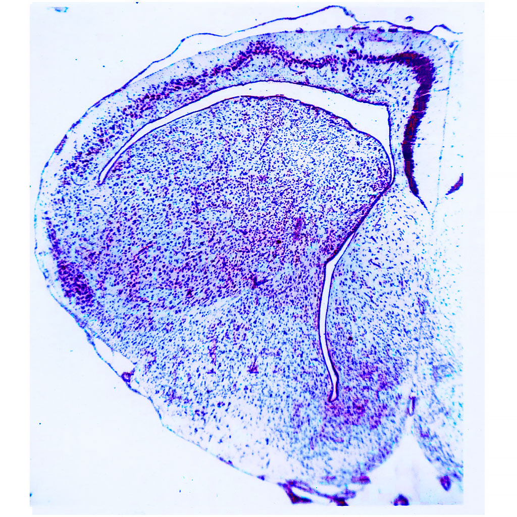
Description: Frontal (transversal) paraffin section through a telencephalic hemisphere of the Siberian sturgeon Acipenser baerii Brandt, 1869 stained with cresyl violet by the Nissl method. The section shows the overall cytoarchitecture of an everted-type hemisphere. The section also clearly shows that fishes have only the median ventricle. The neurons are clustered near the surface of the brain and as they migrate deeper into the brain they form a layered structure in the dorsal regions. In the ventral (subpallial) areas, the neurons are even more concentrated near the median ventricle. This type of brain development complicates the comparison with the brains of other vertebrates and suggests that the CNS of this group of fishes (ray-finned fishes) has evolved along its own distinct path. Organs: сentral nervous system, cerebral brain or its part Methods: Neurohistology - Nissl staining |
|
Series
|

|
|
|
Preparation |
Description: Frontal (transverse) paraffin section of a telencephalic hemisphere of the common frog stained with cresyl violet by the Nissle method. The section shows the overall cytoarchitecture of a hemisphere of an amphibian. The section also clearly shows that the great majority of neurons in both the pallial and subcortical (subpallial) structures are located near the cerebral ventricles. There are only a few neurons inside the wall of the hemisphere (closer to the surface), which indicates a low organizational level of brain structures and the lack of true cortical formations. Organs: сentral nervous system, cerebral brain or its part Methods: Neurohistology - Nissl staining |
|
Series
|
|
|
|
Preparation |
Description: Frontal (transverse) paraffin section through a telencephalic hemisphere of the steppe agama Agama sanguinolenta (Pallas, 1814) stained with cresyl viotet by the Nissl method. The section shows the overall cytoarchitecture of a hemisphere of a lizard. The section also shows how the true cortical structure begins to be formed in the pallial structures (neurons detach from the surface of the ventricle and form a distinct cortical layer in the dorsal regions of the hemisphere). The reptiles are the first vertebrates to form the true cortical plate (the forerunner of the mammalian cortex). Organs: сentral nervous system, cerebral brain or its part Methods: Neurohistology - Nissl staining |
|
Series
|

|
|
|
Preparation |
Description: Frontal (transverse) paraffin sections through a telencephalic hemisphere of a carrion crow stained with toluidine blue by the Nissl method. The section shows the overal structure (cytoarchitecture) of a telencephalic hemisphere, and the distribution pattern of neuronal populations in different regions of the hemisphere. The boundaries between these regions are clearly visible. Organs: сentral nervous system, cerebral brain or its part Methods: Neurohistology - Nissl staining |
|
Series
|

|
|
|
Preparation |
Description: Frontal (transverse) celloidin section through a telencephalic hemisphere of a carrion crow impregnated with silver salts by the Golgi method. Region of the pallium. The section shows a separately stained nerve cell with a system of dendritic (d) and axonal (ax) arborizations. Note a rich network of axonal collaterals (ac) and the lack of spines on the dendrites. According to the classification of E. Ramon-Molinera, these neurons belong to a group of highly specialized, idiodendritic cells, which shows a high level of neuronal organization of the brain in Corvidae. Organs: сentral nervous system, cerebral brain or its part Methods: Neurohistology - Golgi method of silver impregnation of nervous elements |
|
Series
|

|
|
|
Preparation |
Description: Frontal (transverse) celloidin section through a telencephalic hemisphere of the carrion crow Corvus corone L. impregnated with silver salts by the Golgi method. Dorsal region of the pallium (HD – hyperstriatum dorsocellulare). The section shows a group of densely branched, spiny neurons that form the basis of multicellular neuronal groups in the pallium of birds (association). They are viewed as an analogue of modules in the mammalian cerebral cortex. These neurons are classified as specialized Class II allodendritic cells. Organs: сentral nervous system, cerebral brain or its part Methods: Neurohistology - Golgi method of silver impregnation of nervous elements |
|
Series
|

|
|
|
Preparation |
Description: Frontal (transverse) celloidin section through a telencephalic hemisphere of a Japanese quail impregnated with silver salts by the Golgi method (cell bodies and neurites are stained black). Central area of the hemisphere. Silver stained a single neuron and several adjacent glial cells (presumably astrocytes). This neuron is classified as a Class I allodendritic radial neuron. These neurons are considered to be the elements controling intrahemispheral connections, because they have long axons reaching beyond the local cell group and even the local region of the hemisphere. Organs: сentral nervous system, cerebral brain or its part Methods: Neurohistology - Golgi method of silver impregnation of nervous elements |
|
Series
|

|
|
|
Preparation |
Description: Frontal (transverse) celloidin section through a telencephalic hemisphere of the common chaffinch Fringilla coelebs L. impregnated with silver salts by the Golgi method. Dorsal region of the pallium (HD – hyperstriatum dorsocellulare). The section shows a separate short-axon, spineless neuron. This is one of the most highly differentiated neurons in the CNS structures of vertebrates, which belongs to highly specialized idiodendritic cells (by classification of E. Ramon-Molinera). In general, these neurons are found in the pallial regions of the telencephalon in Passeriformes (especially in Corvidae) and mammals (in Carnivora and primates). Organs: сentral nervous system, cerebral brain or its part Methods: Neurohistology - Golgi method of silver impregnation of nervous elements |
|
Series
|

|
|
|
Preparation |
Description: Frontal (transverse) celloidin section through a telencephalic hemisphere of a Japanese quail impregnated with silver salts by the Golgi method. Dorsal region of the pallium (HD – hyperstriatum dorsocellulare) showing a group of neurons of various types that form one of the numerous multineuron groups of the pallium ("nests"). Each nest consists of 3-15 cells of different types positioned a certain distance from one another (in contrast with "associations", where neurons are all closely grouped together and can even form interneuronal soma-somatic contacts). This group includes both densely-branched (1) and sparsely-branched (2) radial neurons that belong to Class II allodendritic cells. The dendrites bear a large number of spines. The section also shows impregnated blood vessels (bv) and deposits of silver salts (black spots). Organs: сentral nervous system, cerebral brain or its part Methods: Neurohistology - Golgi method of silver impregnation of nervous elements |
|
Series
|

|
|
|
Preparation |
Description: Frontal (transverse) celloidin section through a telencephalic hemisphere of a Japanese quail impregnated with silver salts by the Golgi method. Central region of the pallium (M - mesopallium) showing a group of neurons of various types that form one of the numerous multineuron groups of the pallium ("nests"). In addition to the neurons, the multicellular groups include glial cells. The preparation clearly shows several glial cells (presumably the astrocytes - ast). They come in close contact with the neurites and cell bodies of the neurons to form a neuro-glial complex (or using the modern terminology, "the neuro-glial unit"). This emphasizes the important role of glia in the functioning of the nerve centers. The section also shows impregnated blood vessels (bv) and deposits of silver salts (black spots). Organs: сentral nervous system, cerebral brain or its part Methods: Neurohistology - Golgi method of silver impregnation of nervous elements |
|
Series
|

|
|
|
Preparation |
Description: Cytoarchitecture of a part of the paleostriatum (PP) of a crow. One can see large neurons that are surrounded by glial cells (neuroglial comlex), as well as numerous small neurons and glia. A diffuse distribution of neurons is characteristic for this region of a hemisphere. Neuroglial associations are barely present. Organs: сentral nervous system, cerebral brain or its part, neuron(s) Methods: Standard histology - toluidine blue |
|
Series
|

|
|
|
Preparation |
Description: A fragment of the dorsal hyperpallium (HD) of the telencephalon of a hooded crow. The image shows a short-axon aspiny stellate neuron from the HD. Note abundant axonal arborisations (axc) and aspiny dendrites (d). According to the E. Ramon-Moliner classification, this type of neurons belongs to the class of highly specialised idiodendritic neurons. The presence of such neurons in the birds brain indicates extremely high stage of neuronal organization of their pallium. This feature places corvids closer to primates; and dorsal regions of the hemispheres are comparable with cortex formations the mammals. Organs: сentral nervous system, cerebral brain or its part, neuron(s) Methods: Neurohistology - Golgi method of silver impregnation of nervous elements |
|
Series
|

|
|
|
Preparation |
Description: A fragment of the dorsal hyperpallium (HD) of the telencephalon of a chaffinch. The image shows a spiny radial neuron from the dorsal area of the chaffinch hemispheres. According to the E. Ramon-Moliner classification, this type of neurons belongs to specialised allodendritic neurons of the class II. Organs: сentral nervous system, cerebral brain or its part, neuron(s) Methods: Neurohistology - Golgi method of silver impregnation of nervous elements |
|
Series
|

|
|
|
Preparation |
Description: The image shows a fragment of dorsolateral corticoid plate in a hemisphere the crows telencephalon. Large multipolar neurons (mn) do not form any distinct laminated structures. Numerous glial cells (g) and small blood vessels (c) are also visible. The corticoid plate in corvids is weakly developed. Organs: сentral nervous system, cerebral brain or its part, neuron(s) Methods: Standard histology - toluidine blue |
|
Series
|

|
|
|
Preparation |
Description: The image shows a fragment of the neuronal structure of the dorsolateral corticoid plate in the telencephalon. Multipolar spiny radial neurons are visible. The type of dendrite branching of the most neurons suggests that they belong to the Class I radial neurons of the allodendrite type (according to the classification of E. Ramon-Moliner). This indicates low differentiation level of this part of the pallium. According to present views, corticoid plate of the birds telencephalon belongs to the limbic system of the brain. Organs: сentral nervous system, cerebral brain or its part, neuron(s) Methods: Neurohistology - Golgi method of silver impregnation of nervous elements |
|
Series
|

|
|
|
Preparation |
Description: A fragment of the mesopallium (M) - a region in the pallium of the telencephalon of a chaffinch. The image shows a neuron that belongs to radial neurons of the Class I allodendritic type. Such neurons are rare in the pallium. They are characterized by weakly developed branching of the dendrites and small number of spines. Glial cells - astrocytes - are visible among the neurons. Dark spots on the image - unspecific silver sediment. Small blood vessels are also stained. Organs: сentral nervous system, cerebral brain or its part, neuron(s) Methods: Neurohistology - Golgi method of silver impregnation of nervous elements |
|
Series
|
|
|
|
Preparation |
Description: A fragment of the nidopallium (N) - a region of the avian pallium. The image shows a group of neurons and glial cells (association). Neuroglial association provide the basis of the avian pallium. They are analogous to the modular structure of the mammalian telencephalon. Glial cells (astrocytes) lie among the neurons. Rich mesh of small blood vessels is also visible. Organs: сentral nervous system, cerebral brain or its part, neuron(s) Methods: Neurohistology - Golgi method of silver impregnation of nervous elements |
|
Series
|

|
|
|
Preparation |
Description: Frontal (transverse) celloidin section through an area of a telencephalic hemisphere of a rat in the region of the hippocampus impregnated with silver salts by the Golgi method. The section shows a separately stained typical pyramidal neuron in the pyramidal layer of the hippocampus (archicortex). The neuron has a characteristic pattern of the dendritic arborization (the apical dendrite with lateral branches). The dendrites bear a large number of spines. According to the classification of E. Ramon-Molinera, these neurons are classified as highly-specialized idiodendritic cells and are similar to the pyramidal cells of the neocortex. Organs: сentral nervous system, cerebral brain or its part Methods: Neurohistology - Golgi method of silver impregnation of nervous elements |
|
Series
|

|
|
|
Preparation |
Description: Frontal (transverse) paraffin sections through a telencephalic hemisphere of a European hedgehog stained by the Nissl method. The section shows the overall cytoarchitecture of a hemisphere and all major regions of the cortex and some structures of the subcortex. This photograph is of special interest because the insectivores are one of the most ancient orders of the placental mammals. This group shows the initial steps in the evolution of the main cortical regions (archicortex, paleocortex and neocortex). This group is also important because the insectivores are the ancestors of the primates. Organs: сentral nervous system, cerebral brain or its part Methods: Neurohistology - Nissl staining |
|
Series
|

|
|
|
Preparation |
Description: Nervous system of a free-swimming planktotrophic larva. Early pioneer neurons labeling main nerve structures (ventral cords, cerebral commissure, circumoral ring, nerve of the prototroch) and transitory larval structures (apical organ, sensory neurons). Cilia are shown in red, serotonin-containing nerve elements in green. Organs: larval nervous system Methods: Immunohistochemistry - serotonin, |
|
Series
|

|
|
|
Preparation |
Description: Pioneer serotonin-containing neuron of a larva showing the cell body, a long projection with the growth cone and lateral branches. Serotonin in the neuron is shown in green. Organs: larval nervous system Methods: Immunohistochemistry - serotonin, |
|
Series
|

|
|
|
Preparation |
Description: Nervous system of a chiton larva. Sensory serotonin-containing neurons in the episphere. Cilia are show in red, serotonin-containing nerve elements in green. Organs: larval nervous system Methods: Immunohistochemistry - serotonin, |
|
Series
|

|
|
|
Preparation |
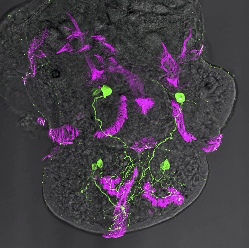
Description: Central ganglia (cerebral, pedal, parietal, and visceral) and mantle innervation in a pond snail veliger. Surface ciliary bunches are visible. Organs: сentral nervous system, cerebral brain or its part, ganglion / ganglia, commissure, larval nervous system Methods: Immunohistochemistry - FMRFamide, |
|
Series
|

|
|
|
Preparation |
Description: Apical organ neurons are located in the center of the episphere. Prototroch cilia are visible along the body edge. Organs: сentral nervous system, cerebral brain or its part, ganglion / ganglia, commissure, larval nervous system Methods: Immunohistochemistry - serotonin, |
|
Series
|
|
|
|
Preparation |
Description: Apical neurons and developing cerebral ganglia at the head region of the larva. Developing pedal ganglia and innervation of foot ciliary structures. Organs: сentral nervous system, cerebral brain or its part, ganglion / ganglia, commissure, larval nervous system Methods: Immunohistochemistry - serotonin, |
|
Series
|
|
|
|
Preparation |
Description: 5HT antibody marks neurons of the apical organ, FMRF-amid antibody - peripheral pioneer neurons, α-tubulin antibody - surface cilia. Organs: larval nervous system Methods: Immunohistochemistry - serotonin, |
|
Series
|

|
|
|
Preparation |
Description: Original image is combined with 3D reconstruction of the apical organ, cerebral ganglia complex and the ventral nerve cords. Organs: larval nervous system Methods: Immunohistochemistry - FMRFamide |
|
Series
|

|
|
|
Preparation |
Description: Serotonin antidody marks nervous elements. Neurons within cerebral and pedal ganglia are visible as well as innervation of mantle complex and foot in Lymnaea veliger. Organs: larval nervous system Methods: Immunohistochemistry - serotonin, |
|
Series
|

|
|
|
Preparation |
Description: Early peripheral pioneer neurons and their processes scaffolding the developing adult central nervous system in early veliger of Lymnaea stagnalis. Organs: larval nervous system Methods: Immunohistochemistry - FMRFamide, |
|
Series
|

|
|
|
Preparation |
Description: FMRF-amide immunostaining of the early sensory cells in the episphere of a larva. Cilia are stained with alpha-tubuline. Organs: neuron(s), receptor cell(s), larval nervous system Methods: Immunohistochemistry - FMRFamide, |
|
Series
|

|
|
|
Preparation |
Description: Three symmetrical cells at the base of each arm and solitary cells along the ventral arm surface are visible. Cell processes run along the arm ciliary bands. Faintly stained neurons are located around the mouth. Organs: larval nervous system Methods: Histochemistry - catecholaminergic nervous elements (GIF) |
|
Series
|

|
|
|
Preparation |
Description: Neurons around the mouth are visible. Organs: larval nervous system Methods: Histochemistry - catecholaminergic nervous elements (GIF) |
|
Series
|

|
|
|
Preparation |
Description: Image shows differentiating neurons of the apical organ and the oral lobe. A solitary neuron is visible in the stomach. Organs: larval nervous system Methods: Immunohistochemistry - serotonin, |
|
Series
|

|
|
|
Preparation |
Description: Developing cerebral ganglion with the ventral nerve cords. Below the ventral cords is a nerve plexus underneath the ventral ciliary band. Ring-shaped lateral nerves innervating the ring-shaped ciliary bands are visible in the body. Cilia are show in red, serotonin-containing nerve elements in green. Organs: сentral nervous system, nerve cord, nerve plexus, cerebral brain or its part Methods: Immunohistochemistry - serotonin, |
|
Series
|

|
|
|
Preparation |
Description: Developing head ganglion and ventral nerve cords. Fibers innervating the muscular bulbus, and local network of the anterior portion of the digestive system are visible. Organs: cerebral brain or its part, nerve cord, nerve plexus, visceral nervous system Methods: Immunohistochemistry - FMRFamide, |
|
Series
|

|
|
|
Preparation |
Description: Cerebral ganglion, ventral nerve cord, circular ciliary bands are seen. Organs: сentral nervous system, nerve cord, nerve plexus, cerebral brain or its part, dorsal (spinal) cord or ventral cord, ganglion / ganglia, connective, commissure, neuron(s), peripheral nervous system Methods: Immunohistochemistry - serotonin, |
|
Series
|

|
|
|
Preparation |
Description: Cerebral ganglion, ventral nerve cord, mouth and circular ciliary bands innervation, as well as peripheral body wall innervation are visible. Alpha-tubulin staining revealed several types of surface cilia: locomotory and sensory. Organs: сentral nervous system, nerve cord, nerve plexus, cerebral brain or its part, dorsal (spinal) cord or ventral cord, ganglion / ganglia, connective, commissure, neuron(s), peripheral nervous system Methods: Immunohistochemistry - serotonin, |
|
Series
|

|
|
|
Preparation |
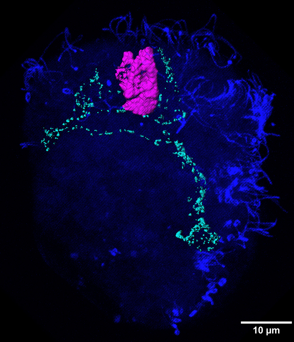
Description: The image shows several groups of neurons that produce ascending and descending connections in the left and right halfs of the supraesophageal ganglion. Organs: сentral nervous system, ganglion / ganglia, nerve, neuron(s) Methods: Retrograde and anterograde tracing techniques, extra- and intracellular labeling of neurons - retro-anterograde transport of nickel chloride method |
|
Series
|
|
|
|
Preparation |
Description: The image shows several groups of neurons that produce ascending and descending connections in the left and right halfs of the supraesophageal ganglion. Organs: сentral nervous system, ganglion / ganglia, nerve, neuron(s) Methods: Retrograde and anterograde tracing techniques, extra- and intracellular labeling of neurons - retro-anterograde transport of nickel chloride method |
|
Series
|

|
|
|
Preparation |
Description: The image shows several groups of neurons that produce ascending and descending connections in the left and right halves of the supraesophageal ganglion. Organs: сentral nervous system, ganglion / ganglia, nerve, neuron(s) Methods: Retrograde and anterograde tracing techniques, extra- and intracellular labeling of neurons - retro-anterograde transport of nickel chloride method |
|
Series
|

|
|
|
Preparation |
Description: The image shows several groups of neurons that produce connections in the left and right halves of the supraesophageal ganglion, and send processes in the right cervical connective. Organs: сentral nervous system, cerebral brain or its part, connective, neuron(s) Methods: Retrograde and anterograde tracing techniques, extra- and intracellular labeling of neurons - retro-anterograde transport of nickel chloride method |
|
Series
|
|
|
|
Preparation |
Description: The image shows several groups of neurons that send processes in the ventral nerve cord postereior to the 1st thoracal ganglion. Organs: сentral nervous system, ganglion / ganglia, nerve, neuron(s) Methods: Retrograde and anterograde tracing techniques, extra- and intracellular labeling of neurons - retro-anterograde transport of nickel chloride method |
|
Series
|

|
|
|
Preparation |
Description: The image shows several groups of neurons that send processes in the right cervical connective. Organs: сentral nervous system, ganglion / ganglia, nerve, neuron(s), neural tract, connective Methods: Retrograde and anterograde tracing techniques, extra- and intracellular labeling of neurons - retro-anterograde transport of nickel chloride method |
|
Series
|

|
|
|
Preparation |
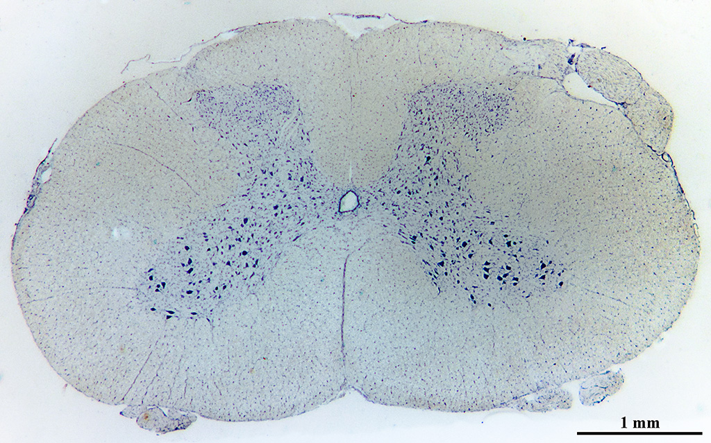
Description: With the help of neurofibril revealing, bodies and processes of neurons were stained. Large multipolar motoneurons are visible in the gray matter. Organs: dorsal (spinal) cord or ventral cord, сentral nervous system, neuron(s) Methods: Neurohistology - Cajal's method of silver impregnation of nervous elements |
|
Series
|

|
|
|
Preparation |
Description: With the help of neurofibril revealing, bodies and processes of neurons were stained. Large multipolar motoneurons in gray matter are visible. Organs: dorsal (spinal) cord or ventral cord, сentral nervous system, neuron(s) Methods: Neurohistology - Cajal's method of silver impregnation of nervous elements |
|
Series
|

|
|
|
Preparation |
Description: With the help of neurofibril revealing, bodies and processes of neurons are stained. Meninges, gray matter and white matter can be seen. Organs: dorsal (spinal) cord or ventral cord, сentral nervous system Methods: Neurohistology - Cajal's method of silver impregnation of nervous elements |
|
Series
|
|
|
|
Preparation |
Description: With the help of a neurofibril revealing method, bodies and processes of neurons were stained. Large multipolar motoneurons are visible in the gray matter. Organs: dorsal (spinal) cord or ventral cord, сentral nervous system, neuron(s) Methods: Neurohistology - Cajal's method of silver impregnation of nervous elements |
|
Series
|

|
|
|
Preparation |
Description: Tigroid substance (endoplasmic reticulum) in neurons in the central channel area of the spinal cord, revealed with Nissl staining. Organs: dorsal (spinal) cord or ventral cord, сentral nervous system, neuron(s) Methods: Neurohistology - Nissl staining |
|
Series
|

|
|
|
Preparation |
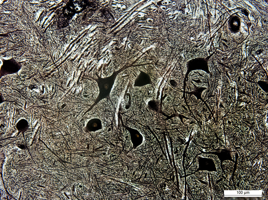
Description: Nissl staining of the tigroid substance (endoplasmic reticulum) in motoneurons and glial cells in the anterior horns of the spinal cord. Organs: dorsal (spinal) cord or ventral cord, сentral nervous system, neuron(s) Methods: Neurohistology - Nissl staining |
|
Series
|

|
|
|
Preparation |
Description: With the help of the Nissl staining, the tigroid substance (endoplasmic reticulum) in motoneurons of the anterior horns of the spinal cord was revealed. Organs: dorsal (spinal) cord or ventral cord, сentral nervous system, neuron(s) Methods: Neurohistology - Nissl staining |
|
Series
|
|
|
|
Preparation |
Description: With the help of the Nissl staining, the tigroid substance (endoplasmic reticulum) in motoneurons of the anterior horns of the spinal cord was revealed. Organs: dorsal (spinal) cord or ventral cord, сentral nervous system Methods: Neurohistology - Nissl staining |
|
Series
|

|
|
|
Preparation |
Description: Nissl staining of the tigroid substance (endoplasmic reticulum) in alfa and gamma motoneurons of the anterior horns of the spinal cord. Organs: dorsal (spinal) cord or ventral cord, сentral nervous system, neuron(s) Methods: Neurohistology - Nissl staining |
|
Series
|

|
|
|
Preparation |
Description: The neural tube and the developing spinal cord are visible. Organs: larval nervous system Methods: Standard histology - hematoxylin, |
|
Series
|

|
|
|
Preparation |
Description: General structure of the cerebellum of a dog. The layered structure of the cerebellum, gray matter and white matter were revealed. Organs: сentral nervous system, neuron(s), cerebral brain or its part Methods: Neurohistology - Cajal's method of silver impregnation of nervous elements |
|
Series
|

|
|
|
Preparation |
Description: General structure of the cerebellum of a dog. The layered structure of the cerebellum was revealed. Organs: сentral nervous system, neuron(s), cerebral brain or its part Methods: Neurohistology - Cajal's method of silver impregnation of nervous elements |
|
Series
|

|
|
|
Preparation |
Description: General structure of a gyrus in the cerebellar cortex of a dog. The cellular composition of the layers of the gray matter is revealed. Organs: сentral nervous system, neuron(s), cerebral brain or its part Methods: Standard histology - hematoxylin, |
|
Series
|

|
|
|
Preparation |
Description: General structure of a gyrus of the cerebellar cortex. The layers in the gray matter and Purkenje cells are visible. Organs: сentral nervous system, neuron(s), cerebral brain or its part Methods: Standard histology - hematoxylin, |
|
Series
|

|
|
|
Preparation |
Description: Nerve cells and nerve plexuses of different types, afferent and efferent processes Organs: ganglion / ganglia, cerebral brain or its part, neuron(s), larval nervous system Methods: Neurohistology - Golgi method of silver impregnation of nervous elements |
|
Series
|

|
|
|
Preparation |
Description: Caudal part of the ventral nerve cord. Ganglia with connectives and nerves are stained. Organs: ganglion / ganglia, сentral nervous system, dorsal (spinal) cord or ventral cord, connective, larval nervous system Methods: Neurohistology - supravital methylene blue staining |
|
Series
|

|
|
|
Preparation |
Description: Region of cell bodies, neuropil, bodies and processes of neurons of the ganglion are stained. One can see connective nerve processes that run peripherally through lateral nerves. Organs: сentral nervous system, dorsal (spinal) cord or ventral cord, ganglion / ganglia, connective, larval nervous system Methods: Neurohistology - supravital methylene blue staining |
|
Series
|

|
|
|
Preparation |
Description: Region of cell bodies, neuropil, bodies and processes of neurons of the ganglion are stained. One can see connective nerve processes that go peripherally through the 1st and 2nd paired abdominal nerves. Organs: сentral nervous system, dorsal (spinal) cord or ventral cord, ganglion / ganglia, connective, larval nervous system Methods: Neurohistology - supravital methylene blue staining |
|
Series
|
|
|
|
Preparation |
Description: Region of cell bodies, neuropil, bodies and processes of neurons of the ganglion are stained Organs: сentral nervous system, dorsal (spinal) cord or ventral cord, ganglion / ganglia, connective, larval nervous system Methods: Neurohistology - supravital methylene blue staining |
|
Series
|

|
|
|
Preparation |
Description: Region of cell bodies, neuropil, bodies and processes of neurons of the ganglion are stained. One can see connective nerve processes that run peripherally through lateral nerves. Organs: сentral nervous system, dorsal (spinal) cord or ventral cord, ganglion / ganglia, connective, larval nervous system Methods: Neurohistology - supravital methylene blue staining |
|
Series
|

|
|
|
Preparation |
Description: Whole mount. Innervation of sensory setae. Organs: nerve, Sensory organ Methods: Neurohistology - supravital methylene blue staining |
|
Series
|

|
|
|
Preparation |
Description: Whole mount. Innervation of sensory setae. Organs: nerve, Sensory organ Methods: Neurohistology - supravital methylene blue staining |
|
Series
|

|
|
|
Preparation |
Description: Whole mount. Innervation of sensory setae. Organs: nerve, Sensory organ Methods: Neurohistology - supravital methylene blue staining |
|
Series
|

|
|
|
Preparation |
Description: Whole mount. Innervation of sensory setae. Organs: nerve, Sensory organ Methods: Neurohistology - supravital methylene blue staining |
|
Series
|

|
|
|
Preparation |
Description: The image shows the neuropil and the neuron somas area, as well as descending pathways, which pass from the anterior connectives through the ganglion. Organs: сentral nervous system, ganglion / ganglia, neuron(s), larval nervous system Methods: Standard histology - hematoxylin, |
|
Series
|

|
|
|
Preparation |
Description: Nervous elements in the metathoracic ganglion of a locust involved in the innervation of the bifunctional muscle 120. Nervous fibers and 4 motoneurons are visible. Organs: сentral nervous system, ganglion / ganglia, neuron(s), nerve, larval nervous system Methods: Retrograde and anterograde tracing techniques, extra- and intracellular labeling of neurons - retro-anterograde transport of cobalt chloride method |
|
Series
|

|
|
|
Preparation |
Description: The image shows neuronal bodies and the ganglion neuropole, which includes regions of сommissural and connective nerve fibers, and terninal branching of nerve processes of lateral nerves. Organs: сentral nervous system, nerve plexus, dorsal (spinal) cord or ventral cord, ganglion / ganglia, nerve, neuron(s), larval nervous system, visceral nervous system Methods: Neurohistology - supravital methylene blue staining |
|
Series
|

|
|
|
Preparation |
Description: The image shows terninal branching of nerve processes, сommissural and connective nerve fibers. Organs: сentral nervous system, nerve plexus, dorsal (spinal) cord or ventral cord, ganglion / ganglia, nerve, neuron(s), larval nervous system, visceral nervous system Methods: Neurohistology - supravital methylene blue staining |
|
Series
|

|
|
|
Preparation |
Description: In the caudal part of the ganglion there are numerous branching dendrites of motoneurons that form paired motor nuclei. In the anterior part of the ganglion there are motoneuron fibers that form anterior motor crossing. The unpaired nerve of the vegetative nervous system enters the ganglion caudally, in the median part. Organs: сentral nervous system, nerve plexus, dorsal (spinal) cord or ventral cord, ganglion / ganglia, nerve, neuron(s), larval nervous system, visceral nervous system Methods: Neurohistology - supravital methylene blue staining |
|