
Optical
|
||
|
Preparation Storage |
Collection: Brain of the amphibians (Amphibia) Species: Rana temporaria Linnaeus, 1758 Amphibia Description: Frontal (transverse) paraffin section of a telencephalic hemisphere of the common frog stained with cresyl violet by the Nissle method. The section shows the overall cytoarchitecture of a hemisphere of an amphibian. The section also clearly shows that the great majority of neurons in both the pallial and subcortical (subpallial) structures are located near the cerebral ventricles. There are only a few neurons inside the wall of the hemisphere (closer to the surface), which indicates a low organizational level of brain structures and the lack of true cortical formations. Comments: none Organs: сentral nervous system, cerebral brain or its part Methods: Neurohistology - Nissl staining Publications: none Microscope: Leica DM 6000 with DFC 5000 EC3 camera |
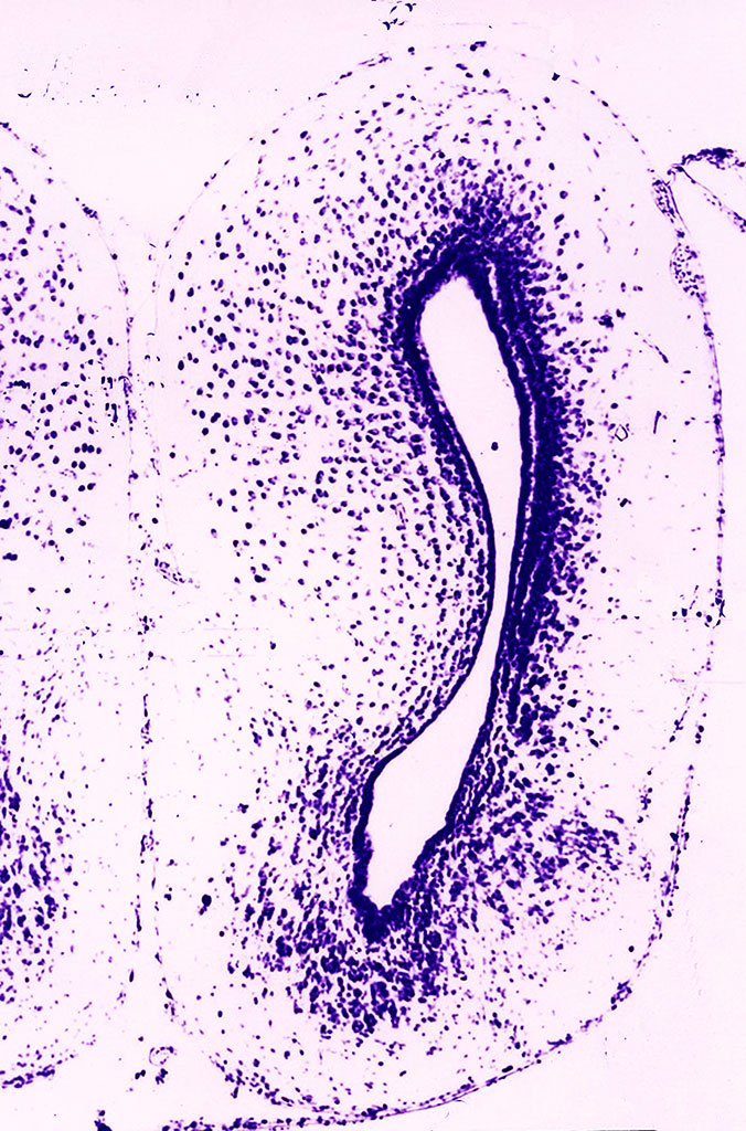
Photo: D.K. Obukhov |
All
|
|
|