
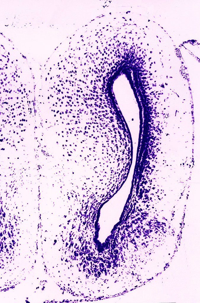
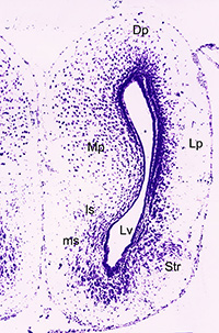
ImageFrontal (transverse) paraffin section through a telencephalic hemisphere of the common frog Rana temporaria Linnaeus, 1758 stained with cresyl violet by the Nissl method |
||
|
← Click to enlarge image Image Type: Single photo Projections Plane: non Color Channels:
Violet — Nissl substance in neurons License: Open with author and institution Comments: none |
|
Image
|
Frontal (transverse) paraffin section of a telencephalic hemisphere of a common frog stained with cresyl violet by the Nissl method. The section shows the overall cytoarchitecture of a hemisphere of an amphibian. The section also shows that the great majority of neurons in both the pallial and subcortical (subpallial) structures are located near the cerebral ventricles. There are only a few neurons inside the wall of the hemisphere (closer to the surface), which indicates a low organizational level of brain structures and the lack of true cortical formations. |
|
Annotated
|
|
|