
General characteristic of the phylum Annelida |
||
|
Author: V.V. Starunov Editor: O.V. Zaitseva Translation |
Annelids or ringed (segmented) worms (phylum Annelida) are a large phylum of archaeostomatous invertebrate with approximately 18,000 species. Their size varies from 0.3 mm (Diurodrilus sp.) to 3 metres (Eunice aphroditois) in length, but in general annelids have small or medium size (from 1 to 10 cm in length). Segmented worms live mostly in sea biotopes, although many species inhabit fresh water (oligochaetes, leeches) and soil (earthworms). |
Content
“
Annelid's body is divided into prostomium (head), a number of body segments, and pygidium (anal lobe).
“
Each side of body segments has locomotory appendages called parapodia. They bear setae (chaeta) and serve for locomotion.
“
The growth zone, which is producing new body segments in front from itself, is located before an anal lobe.
Representative species of annelids (polychaetes)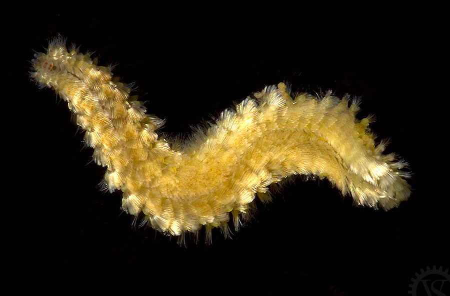
Chrysopetalum occidentale Johnson, 1897 (family Chrysopetalidae). Photo by V.V. Starunov

Syllis sp. (family Syllidae). Photo by V.V. Starunov
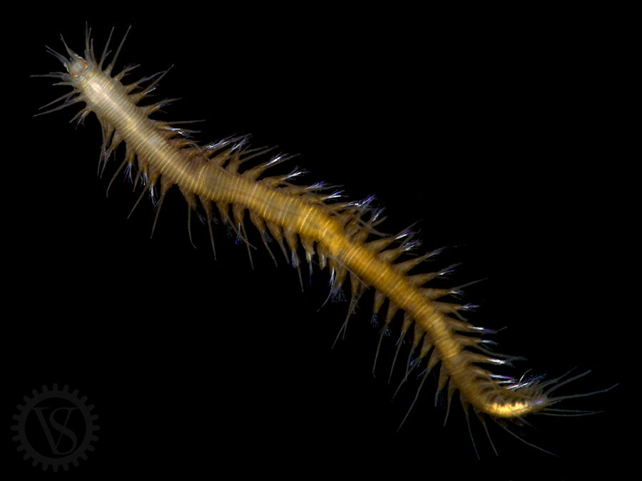
Oxydromus pugettensis (Johnson, 1901) (family Hesionidae). Photo by V.V. Starunov

Phyllodoce sp. (family Phyllodocidae). Photo by V.V. Starunov
|
Systematics of the Annelida |
Systematics of the Annelida has been dramatically changed recently. According to traditional views, all segmented worms were divided into two subtypes: Clitellata and Aclitellata. Cletellates consisted of the class Polychaeta, and Oligochaeta and leeches-like worms (Hirudinea) were included to Aclitellata group. Development of molecular phylogenetic analysis has provoked new research circle in regard to taxonomy of the segmented worms recently. Current system is very different from traditional. For example, according to new data, Oligochaeta and Hirudinea turn to be one subordinate taxon within class Polychaeta. At the same time, taxons, which previously regarded as the separate families within class Polychaeta, became beyond this class. Now these taxons (for example, Oweniidae, Chaetopteridae and Mageloniidae) regard as sister groups. Our site currently uses the traditional variant of annelida classification with dividing into three classes: Polychaeta, Olygochaeta, and Hirudinea. |
|
Body segments |
The incredible morphological diversity is the important feature of annelida. In particular, this diversity relates to neural system structure which demonstrates a high level of evolutionary plasticity. Annelids are extremely heterogeneous by their morphological diversity. Nevertheless, the most of current members of this phylum has particular set of characteristics, and a segmental organization is the most important characteristic from this set. It is possible to divide the whole annelid's body into three main regions: prostomium, body segments, and pygidium (anal lobe). Prostomium contains cerebral ganglion or head brain. It also can have a number of sense organs, such as eyes, antennae, palpus, and nuchal organs. Body segments are the main structural units of the annelid's body. The number of segments can be very variable in different annelid groups. It can be uncertain and dependent only on animal size and age or fixed strongly. Usually the first body segment, which is called peristomium, is different from the next segments by its structure. It has mouth and, in addition, it can bear sensory appendages, or peristomial cirri, which play mainly sensory role. Each side of body segments has locomotory appendages called parapodia. They bear setae (chaeta), and serve for locomotion. Parapodia's and chaeta's structure is species-specific and often used for definition of segmented worms. Each segment can bear only one pair of parapodia. Parapodia of some annelids are reduced to parapodial ridges, and small outgrowths with setae. Parapodia of Oligochaeta and Hirudinea are completely reduced. Members of Oligochaeta have only small amount of chaeta on the lateral side of segments. Chaeta of the most members of Hirudinea are completely reduced too. Segments in the back end of the body are usually smaller than in the middle of the body. This is due mainly to the fact that growth zone, which is producing new body segments in front from itself, is located before the anal lobe. Therefore, the front segments are the oldest, and the back segments are the youngest in the worm's body. The anal lobe, or pigidium, is located in the back end. It bears anus and can have different sensory appendages (pigidial cirrus) which are developed mostly in errant species leading a free-living mode of life. |
|
Body wall |
Annelid's body is covered with cuticle, which is made of collagen fibers. These fibers are disposed by layers, which are perpendicular to each other, giving a characteristic iridescent color to the worm. Usually collagen fibers are located at an angle of 45 degrees to the anteroposterior axis. Such position is the best for providing both strength and elasticity of the integument. The cuticle is secreted by cells of surface epithelium which additionally contains a number of glandular cells secreting mucus. The members of some families contain specialized cells, and with help of these cells animals build protective tube around themselves. Muscle layers are located under the surface epithelium. Annelida's muscle organization is extremely variable and depended on particular animal's mode of life. The body wall muscular system of worms who live in soil is well developed. It consists of outer layer of circular muscle and inner layer of lateral muscle, which usually is divided into four well-distinguished bands. Sometimes a layer of diagonal muscle can be disposed between circular and lateral muscles. Annelids who inhabit a ground surface possess poorly developed circular muscle or even don't have it at all. The whole dermomuscular tube consists of set of different muscular fascicles. Apart from body wall muscles, there is a rather sophisticated complex of muscles which operate parapodia and chaeta, as well as muscles of dissepiments, oblique, and dorsal-ventral muscles. |
|
Coelom |
Coelom is a body cavity which is disposed between body wall and intestine in most of annelids. Each segment has a pair of coelomic compartments (left and right). Septums between left and right coelomic compartments are called mesenteries. Septums between cavities in adjacent segments are called dissepiments. Coelomes are filled by coelomical fluid. Often it is possible to find coelomic cell elements, that are called coelomocytes, inside coelomic cavity. Coelom's functions are various. Coelom is a part of locomotor system of amimal, and plays a role of hydrostatic skeleton. Coelomic fluid can duplicate or even take the whole function of circulatory system consisting in distribution of oxygen and nutrients. In addition, excretory organs are located inside coelomic cavities, and reproductive product development occurs inside these cavities too. |
|
Circulatory and
|
Well-developed closed circulatory system occurs in many members of annelids. The two main vessels are the dorsal and the ventral blood vessels. They are located in dorsal and ventral mesenteries, relatively. Blood is circulated through the dorsal vessel to the front of the body, and through the ventral vessel to the back of the body. Beside this, there are lateral vessels that go through the dissepimens and peri-enteric plexus. The role of propulsion organ is played usually by dorsal vessel or some lateral vessels. Excretory system is represented by paired nephridiums located in each segment. |
|
Digestive system |
Annelid's digestive system goes from the mouth to the anus. Usually it is possible to divide the digestive tract into several parts: pharynx, esophagus, intestine and rectum. Some annelids species have pharynx that can be turn inside out partially or entirely for food capture. Furthermore, pharynx often can have chitinous jaws. |
|
Nervous system |
Annelid's central nervous system consists of brain (cerebral ganglion), circumpharyngeal connectives and abdominal brain (ventral nerve trunk). Peripheral nervous system includes nerves and nerve plexuses inside the body wall, as well as the stomatogastric nervous system. Regardless of general structural plan, the structure of nervous system of various members of polychaete can be substantial variable. The differences relate to all regions of ventral nerve trunk: ganglions, connectives, commissures, segmental and lateral nerves. Cerebral ganglion is located in prostomium. In general it consists of two bilaterally symmetrical parts. Connection between right and left parts occurs through a quantity of commissures. It is possible to distinguish the two main commissures that connect circumpharyngeal connective roots and occur in most of annelids. Tentatively, the annelid's head brain can be divided into three compartments. The front compartment is connected with palpes, the middle one is connected with antennae and eyes, and the back one is connected with nuchal organs. Furthermore, it can be possible to distinguish up to 20 pairs of different cell agglomerations named brain nuclei inside head brain of different members of annelida. The degree of head brain development can be very various depend on mode of life. Sedentary polychaets, who lead inactive mode of life and live in their own tubes all their life, have least developed head brain, which is only paired ganglion without any compartments or nucleus. Burrowing annelids have slightly more developed brain. The most developed brain is found in errant species, who lead free-living mode of life and possess well-developed sense organs. Head brain is connected with abdominal brain by pair of circumpharyngeal connectives. Abdominal brain is usually located on ventral side of animal along the median line. It consists of several pairs of lateral nerve trunks and paired ganglia located in each segment along these nerve trunks. Ganglia in abdominal brain are connected by commissures. The number of commissures is varied from one to seven in different members. Only in few cases (families Nerillidae and Protodrillidae) the number of commissures can be irregular and uncertain. The number of lateral nerve trunks is also uncertain and can be from two to five. Ganglia of ventral nerve cord usually are well distinguished. Only some primitively organized polychaetes don't have clear boundaries between ganglia in the next segments. Ganglia consist of central fibrous area containing the significant amount of synaptic terminals (neuropil), and peripheral area containing cell bodies of neurons with nuclei. Ganglia of the front body segments are fused forming large subpharingeal ganglia. Furthermore, pair ganglia inside one segment can be fused partially or entirely. Fusion of ganglia in the last body segments in the area of caudal sucker is also observed in leeches. Finally, the whole ventral nerve trunk is fused into single nerve centre in Myzostomida. Segmental nerves, whose number varies wildly, derivate from abdominal brain ganglia, and provide innervation of body wall and parapodia. It is possible to distinguish a parapodial nerve, which is larger then other nerves, in species with prominent parapodia. The parapodial nerve often bears a small constellation of nerve cells named parapodial ganglion. Besides segmental nerves, there can be peripheral lateral nerves inside body wall whose number is various in different families. Combined with segmental nerves, they can form regular array of peripheral nerves. This array is similar to orthogon of flatworms. However, this similarity is secondary, because this structure is entirely functional and related to evolving of peripheral regions of nervous system. Stomatogastric nervous system is the important region of peripheral nervous system. It is an assembly of pharynx's and intestine's nerves, ganglia, and neuroplexes, and it is connected with central nervous system by paired stomatogastrical nerves going from head brain and from subpharingeal ganglion, and probably due to segmental nerves going to dorsal side and getting through mesentery to intestine. Detailed structure of annelid's stomatogastric nervous system has been examined shortly and in few species. |
“
Annelid's central nervous system consists of brain (cerebral ganglion), circumpharyngeal connective, and abdominal brain (ventral nerve trunk).
“
Tentatively, the annelid's head brain can be divided into three compartments. The front compartment is connected with palpes, the middle one is connected with antennae and eyes, and the back one is connected with nuchal organs.
“
Abdominal brain consists of several pairs of lateral nerve trunks and paired ganglia located in each segment along these nerve trunks.
“
Annelid's ganglia consist of central fibrous part containing the significant amount of synaptic terminals (neuropil), and peripheral part containing cell bodies of neurons with nuclei.
“
Peripheral nervous system includes nerves and nerve plexuses inside the body wall, as well as stomatogastric nervous system.
|
Sensory organs |
Annelid's sensory structures are various. They include both photo-, chemo- and mechanoreceptors, and multicellular sensory organs. The main part of receptor structures is concentrated in the front end of the body. There are also sensory organs located at each segment and on pigidium. A great amount of mechanoreceptor and chemoreceptor cells is concentrated on sensory appendages: antennae, peristomial, parapodial and pigidial cirri, and palpus. These cells are usually bipolar. Sensory projection of each cell is included in epithelium. Usually it bears sensory cilium on distal end. Axonal projection, that can be significant length, goes to neurons of central neural system. Sensory cells can be arranged into clasters inside specialized sensory ciliated organs. Usually these clasters are considered as chemoreceptor organs. They include nuchal organs, that are located on prostomium, as well as dorsal and lateral ciliated organs, that are located on body segments. Many annelids have one or several pairs of eyes on prostomium. These cerebral eyes are extremely diverse by their structure. The members of plankton polychaets from family Alciopodae have eyes containing light-collecting lens and occupying the most area of prostomium. Eyes with simple structure are inverse. Large eyes are usually converse. Eyes can be located not only on prostomium. The members of several families have eyes that are located on body segments and even on pigidium. Furthermore, single photoreceptors and photoreceptor-like cells can be contained in surface epithelium of body wall. The members of certain annelida families have statocysts that are organs being responsible for sense of gravity. Statosyst is epidermal vesicle with glandular and sensory cells. There are several statolithes inside this vesicle. These sensory organs are particularly typical of borrowing animals. |
“
Annelid's sensory structures include both photo-, chemo- and mechanoreceptors, and multicellular sensory organs.
“
Multicellular sensory ciliated organs include nuchal organs, that are located on prostomium, as well as dorsal and lateral ciliated organs, that are located on body segments.
“
Many annelids have one or several pairs of cerebral eyes on prostomium. Furthermore, the members of several families have eyes that are located on body segments and even on pigidium.
“
Most of the annelids, with the exception of oligochaeta and leeches, are diclinous. Fertilization is external in the most cases.
“
Annelid's cleavage is spiral. Usually the type of gastrulation is epiboly or invagination. Most of ring worms have larvae which is called trochophora and lives as plankton.
“
Certain polychaeta species (for example, Platynereis dumerilii and Capitella teleta) are model objects for developmental biology.
|
Reproductive system |
Most of annelids, with the exception of oligochaeta and leeches, are diclinous. Some species are characterised by epitoky which is the worm's body transformation occuring before reproduction and serving for life in pelagic zone. In some cases epitokal transformation occurs only in the back region of the body. This region grows new head, separates from mother's body and independently swims disseminating reproductive products. Fertilization is external in the most cases. Sometimes the cases of parental care are observed. For example, some members of leeches and some polychaetes of family Syllidae carry juveniles on the dorsal body side. |
|
Ontogeny |
Annelid's cleavage is spiral. Usually the type of gastrulation is epiboly or invagination. Most of ring worms have larvae which is called trochophora and lives as plankton. Then larvae is being segmented, and becoming metatrochophora. During next transformational changes metatrochophora evolves into nektochaeta which is vaguely resembles adult worm with reduced number of segments. Further transformation is related mainly with activation of growth zone, increase of number of body segments, and final transformation of head and pigidium. |
|
Conclusion |
The members of polychaetes hold a special place in collections whose images are represented on our site. At the current moment polychaetes regard as one of the clue groups on the phylogenetic tree of the animal world. According to findings of the latest investigations in molecular biology, it is possible to suggest that polychaeta-like animals (Urbilateria) can be a common ancestor for all bilaterally symmetrical animals. Therefore, ring worm's neural system can have features of primitive organization that is characteristic for the last common ancestor of all the others bilaterally symmetrical animals. It should be noted also that at this date certain polychaeta species, such as Platynereis dumerilii or Capitella teleta, have become one of the routine model objects for different fields of biology, and first of all for developmental biology. |
|
Recommended literature |
|
|