
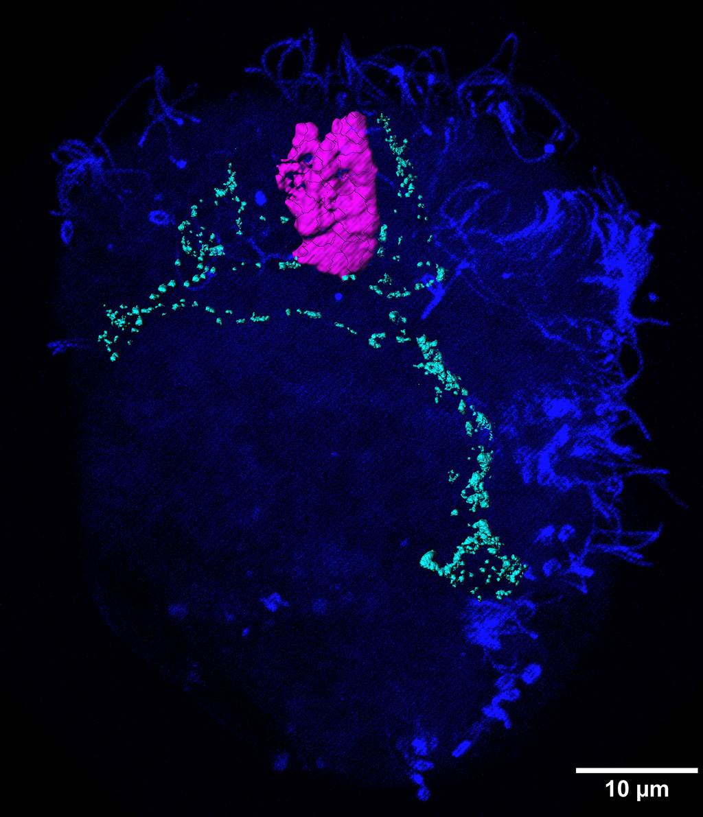
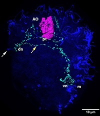
ImageEarly larval neurons of an oyster veliger Crassostrea gigas (Mollusca, Bivalvia). |
||
|
← Click to enlarge image Image Type: Maximum intensity projection Projections Plane: xyz Color Channels:
Violet — serotonin License: Open with author and institution Comments: 3D reconstruction of the apical organ (magenta). |
|
Image
|
5HT antibody marks neurons of the apical organ, FMRF-amid antibody - peripheral pioneer neurons, α-tubulin antibody - surface cilia. |
|
Annotated
|
|
|
© Laboratory of Evolutionary Morphology (Zoological Institute RAS), IDB RAS, SPBU