
Early larval neurons of an oyster veliger Crassostrea gigas (Mollusca, Bivalvia). Annotated Image |
||
|
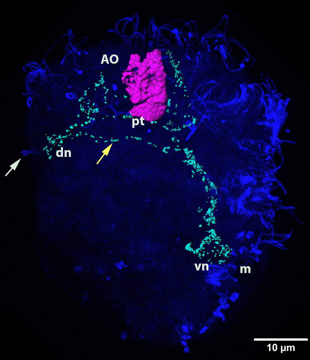
← Click to enlarge image Authors: E.E. Voronezhskaya, V.A. Dyachuk Color Channels:
Violet — serotonin Notation: AO - apical organ; |
|
Image
|
5HT antibody marks neurons of the apical organ, FMRF-amid antibody - peripheral pioneer neurons, α-tubulin antibody - surface cilia. |
|
© Laboratory of Evolutionary Morphology (Zoological Institute RAS), IDB RAS, SPBU