
Brain of fishes (Pisces) |
||
|
19
|
The collection contains permanent histological preparations of the brain of several species of cartilaginous and bony fishes stained using classical neurohistological techniques: Nissl staining and Golgi impregnation. The preparations of each species (several dozens of microscope slides) are stored in separate numbered slide boxes in a special storage cabinet located at the Department of Cytology and Histology of the Saint-Petersburg State University. Each slide is labelled with all essential identifying information. This collection is intended for those students, scientists and teachers who want to learn more about the architecture of the brain and especially its principal region, telencephalon, in several representatives of fishes. The collection is a visual study guide for the teaching courses on zoology, physiology, higher functions of the nervous system, histology, cytology, evolutionary theory, etc., taught at the departments and schools of biological sciences in the universities. |
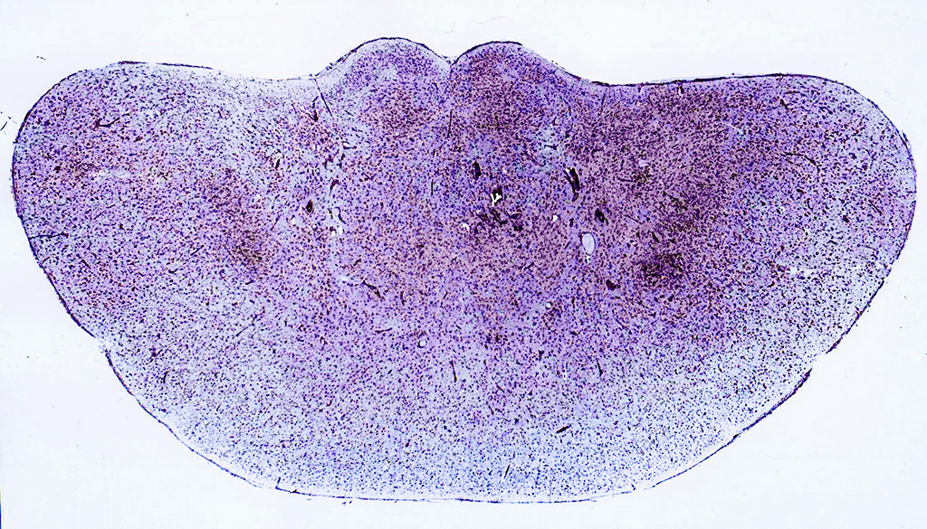
Transverse section through the telencephalic hemispheres of the thornback ray Raja clavata Linnaeus, 1758 stained with cresyl violet by the Nissl method.
|
|
|
Collection author(s): D.K. Obukhov Place of storage: Saint Petersburg State University Taxa: Class Сartilaginous fishes (Chondrichthyes)
Class Bony fishes (Osteichthyes)
Superorder Bony fishes ( Teleostei)
Order Cypriniformes
|
|
Preparations |
||
Preparation SPSU-ODK-PIS-2-1Short axon, aspine, stellate neuron dorsal area telencephalon of a shark Squalus acanthias Linnaeus, 1758 (Chordata, Chondrichthyes, Selachii) |
||
|
Species Squalus acanthias Linnaeus, 1758 Taxon |
Description: Frontal (transverse) celloidin section through an area of a telencephalic hemisphere of the spiny dogfish Squalus acanthias Linnaeus, 1758 impregnated with silver salts by the Golgi method. Staining: Golgi method of silver impregnation of nervous elements |
|
Preparation SPSU-ODK-PIS-3-1Cross section through a telencephalic hemisphere of the Siberian sturgeon Acipenser baerii Brandt, 1869 (Chordata, Osteichthyes, Acipenseriformes) |
||
|
Species Acipenser baerii Brandt, 1869 Taxon |
Description: Frontal (transverse) paraffin section through a telencephalic hemisphere of the Siberian sturgeon Acipenser baerii Brandt, 1869 stained with cresyl violet by the Nissl method. Staining: Nissl staining |
|
")
 paraffin section through a telencephalic hemisphere of the Siberian sturgeon Acipenser baerii Brandt, 1869 stained with cresyl violet by the Nissl method")