
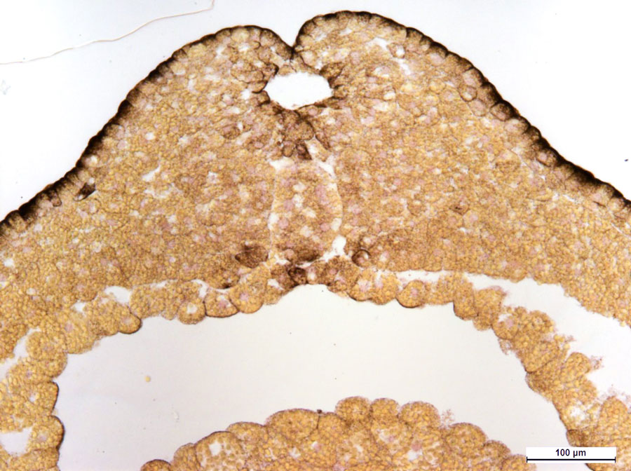
Early neurula (stage of the neural tube formation) of a frog (cross section). Staining with hematoxylin-picrofuchsin. Photo by O.V. Zaitseva and A.N. Shumeev.
|
Welcome to the section devoted to the development of the nervous and sensory systems in ontogeny! The nervous system and sensory organs of the adult animals are not formed all at once during the animal’s development, but together with other functional systems, organs and body parts pass through embryonic, larval (in some representatives of both invertebrate and vertebrate animals) and postembryonic stages. In the animals that have a complex life cycle with alternation of several life forms, as in some insect species and in many parasitic invertebrates, the nervous system may undergo several complex transformations in the course of their development. In this section of the site, you will learn about the general principles and patterns of development of the nervous system and sensory organs during embryonic, larval and juvenile stages of vertebrate and invertebrate animals. The development of any multicellular animal begins when a fertilized egg divides to produce several blastomeres. The development then progresses to the blastula stage (formation of a one-layered embryo), followed by the gastrula stage (formation of the major germ layers), then the formation of the main body axes and organ primordia (definitive or larval, if the organism passes through the larval stage) and finally the formation of the main tissues and organs. Embryonic development ends with the birth (hatching from an egg or an egg mass) of a juvenile or a free-living larva (if this stage is present). In some animals, the larval stages develop inside the egg mass. The transition from the larva to the adult form is accompanied by metamorphosis, during which the larval (temporary, or provisional) organs are partially or completely destroyed and replaced with new (definitive) organs of the adult animal. The organism hatches from the egg or egg mass and continues its life as a juvenile, but the process of formation of most organs and tissues does not stop there and continues also throughout the juvenile stage. In the animals that pass through the larval stage the break down of the provisional organs and development of the definitive organs (including the organs of the nervous system) occur gradually during the larval development or in a catastrophic fashion at the end of the larval stage. The more complex the structure and the larger the body size of the adult, the longer it takes for the animal to complete the juvenile stage, during which the organism finishes the formation of its body, organs and functional systems. In the course of development, different body regions, organs and functional systems emerge at different points in time and develop at various rates (allometric growth). The first organs to appear are those that are the most important at that particular stage and those that require more time to develop due to their complex structure. One of the major of those slowly-developing complex systems is the nervous system. In vertebrates the redistribution of the cellular material leading to the formation of the anlage of the future neural tube occurs as early as the unicellular zygote stage. In invertebrates the first nerve elements (intraepithelial primary receptor cells) appear as soon as the first epithelial layers were formed. Due to the early development of the main nerve centers, the primordia of the central nervous system and sensory organs in juvenile animals are almost always disproportionally large relative to the rest of the body and other organs. |

Cross section through the head of a rabbit embryo. Staining with hematoxylin-eosin. Development of optic brain vesicles and olfactory organ. Photo by O.V. Zaitseva and A.N. Shumeev.

Pilidium pyramidale, a live larva of a White Sea nemertean. Photo by L.P. Flyachinskaya.
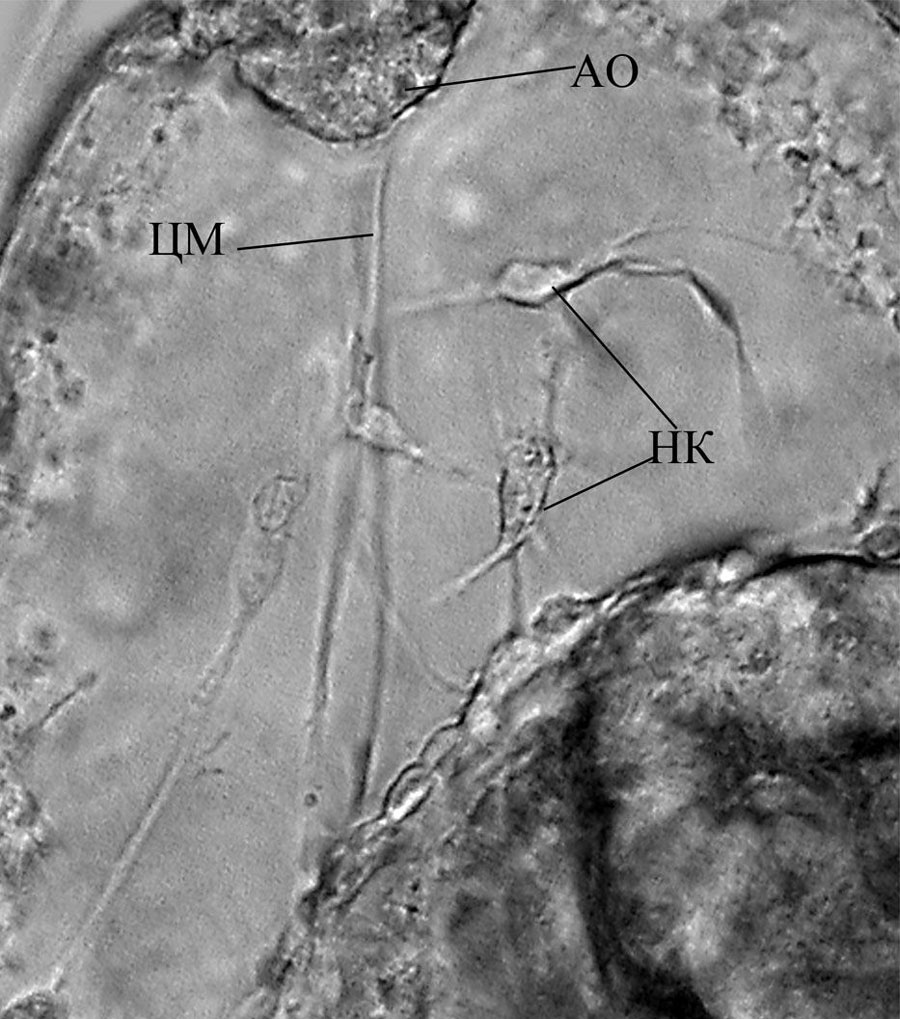
A portion of the body of Pilidium pyramidale, a nemertean live larva with nerve cells (НК) and an apical organ (АО). From: O.V. Zaitseva and L.P. Flyachinskaya (2010).
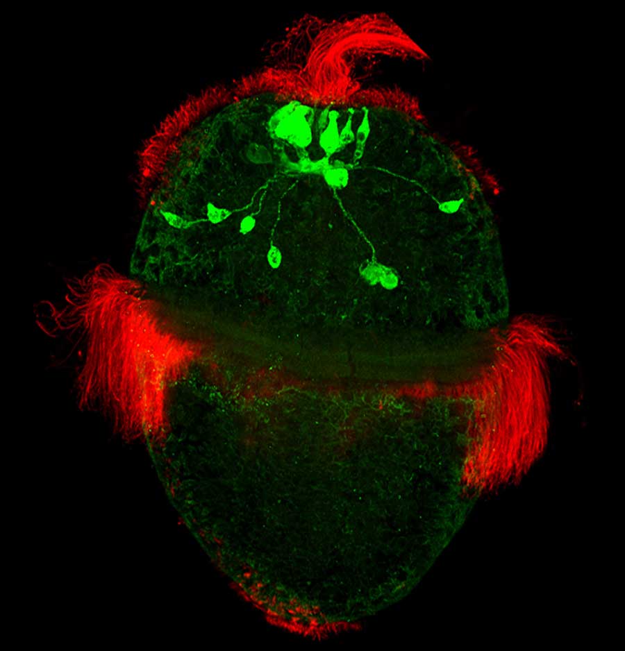
A larva of the chiton Ischnochiton hakodadensis. Serotonin-containing nerve cells (green) and epidermal cilia (red). Photo by L.P. Nezlin.
|
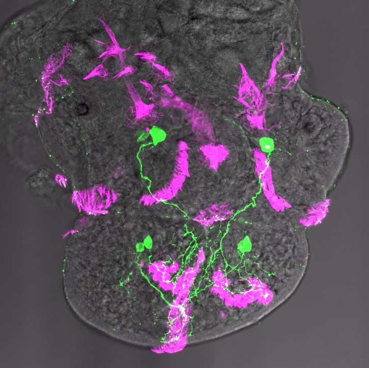
A larva of the mollusc Helisoma trivolvis. Immunohistochemical staining of serotonin-containing nerve cells (green) and epidermal ciliary structures (purple). Photo by E.E. Voronezhskaya.
|
||
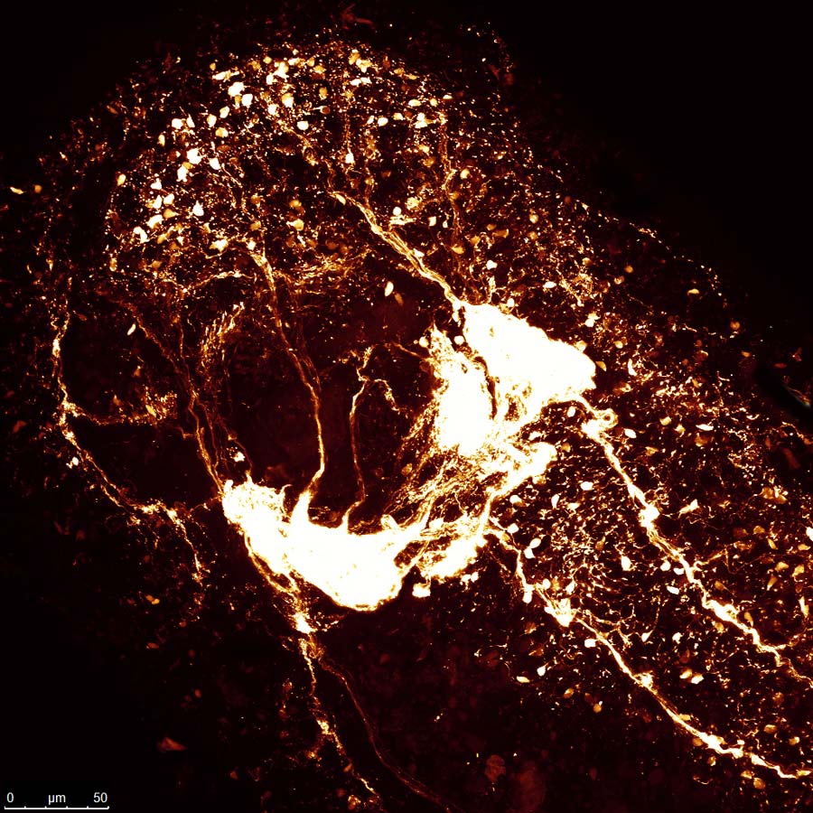
Central and peripheral nervous systems in the anterior body region of a three-month-old individual of the mollusc Cadlina laevis. Histochemical staining of the catecholamine-containing neurons (golden white). Photo by O.V. Zaitseva and A.N. Shumeev.
|
||
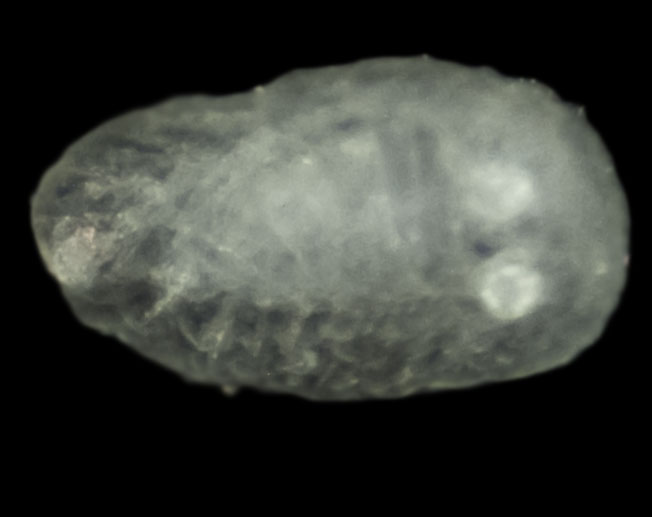
General view of a juvenile of the mollusc Cadlina laevis. Photo by O.V. Zaitseva and A.N. Shumeev.
|
||
© Laboratory of Evolutionary Morphology (Zoological Institute RAS), IDB RAS, SPBU |
||