
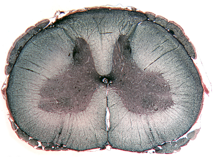
Cross section through the spinal cord of a dog. Cajal's silver impregnation of neurons and neurites. Photo by A.N. Shumeev and O.V. Zaitseva.
|
Welcome to the Methods of Neuromorphology Research! In this section of the site, you will learn the principal techniques employed by scientists to study the structure of the nervous system at various tiers of organisation: gross anatomical, histological, systemic (including the architecture of various nerve nets and their interactions), cellular, subcellular and chemical. You will learn, how the methods should be selected to address specific goals of neuromorphological studies, what advantages or disadvantages are associated with particular methods and what benefits can be gained from their combined application. Our understanding of the architecture of the nervous system is contingent upon the development of new methods and the improvement of classical neuromorphological techniques, and is also directly associated with the development of the microscopy tools. All main methods for studying the morphology of the nervous system can be subdivided into several groups: vital, histological, neurohistological, histochemical, immunohistochemical, methods of retrograde and anterograde transport, methods of experimental degeneration of neurons and their neurites, and transmission and scanning electron microscopy. The synthetic processes in neurons and development of neural structures were actively studied by the autoradiography method based on detection of radioactively labelled isotopes injected into the animals. Today, however, this technique is rarely used. The nervous structures are examined using optical, phase-contrast, fluorescent, electron and confocal laser scanning and multiphoton microscopy. The confocal and multiphoton microscopy together with the development of immunohistochemical markers have opened a new era in the study of the nervous system allowing full 3D reconstruction of nerve nets based on series of optical sections and identification of specific neuroactive substances in the constituent nervous elements. |
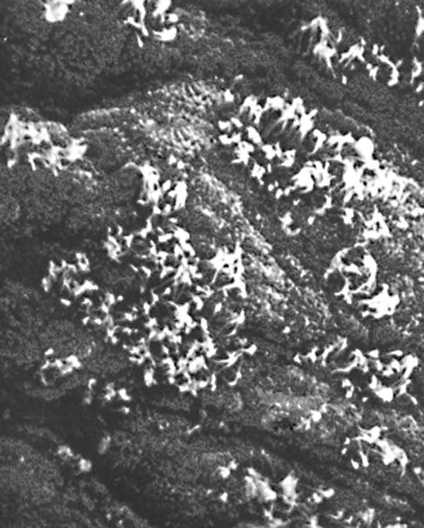
Receptor epithelium of an olfactory tentacle of the mollusc Buccinun undatum. Scanning electron microscopy. Photo by O.V. Zaitseva.
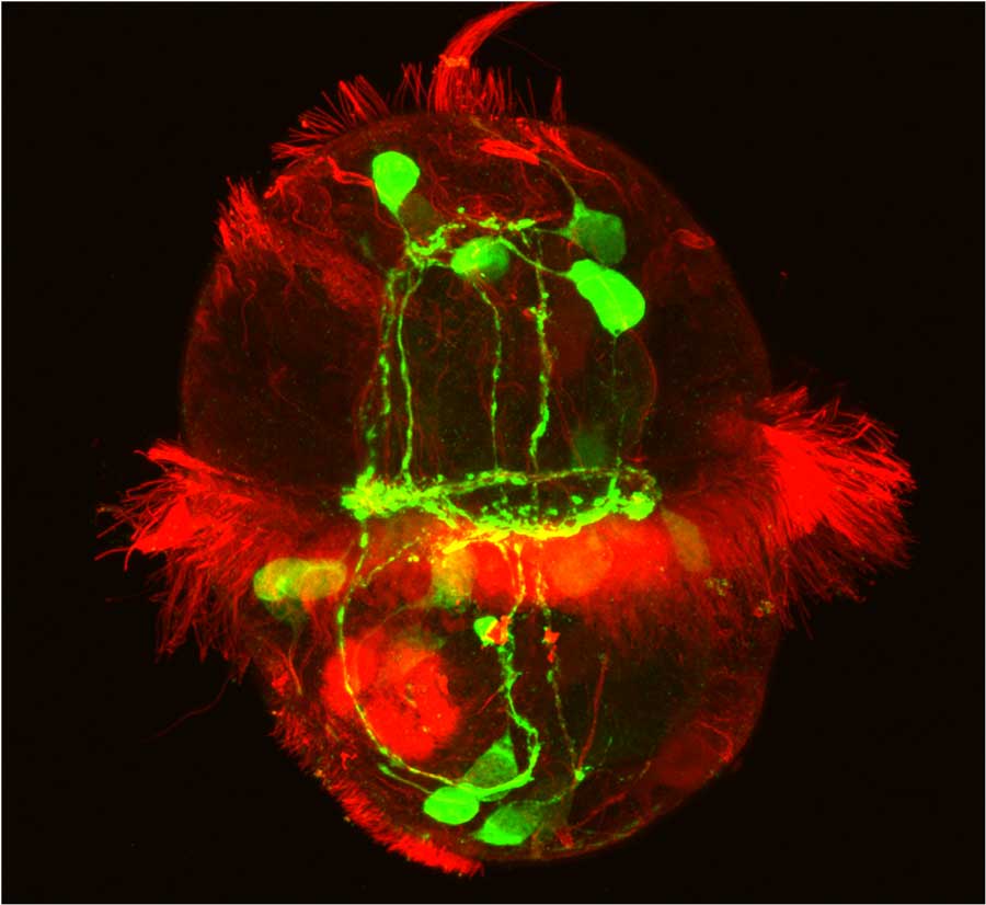
Serotonin-immunoreactive nervous system (green) of a larva of the polychete Phyllodoce maculata. Immunohistochemistry of serotonin and tubulin (red). Photo by L.P. Nezlin.
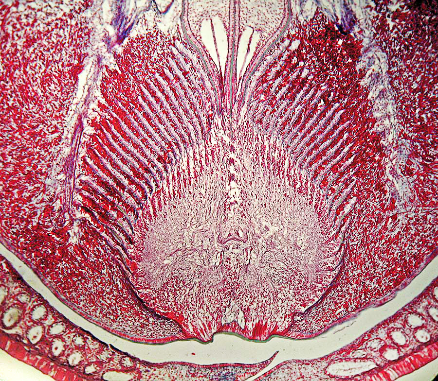
Cross seciton through the brain of the vestimentiferan Riftia pachyptila. Heidenhain's azan staining. Photo by A.V. Ivanov and R.V. Selivanova.
|
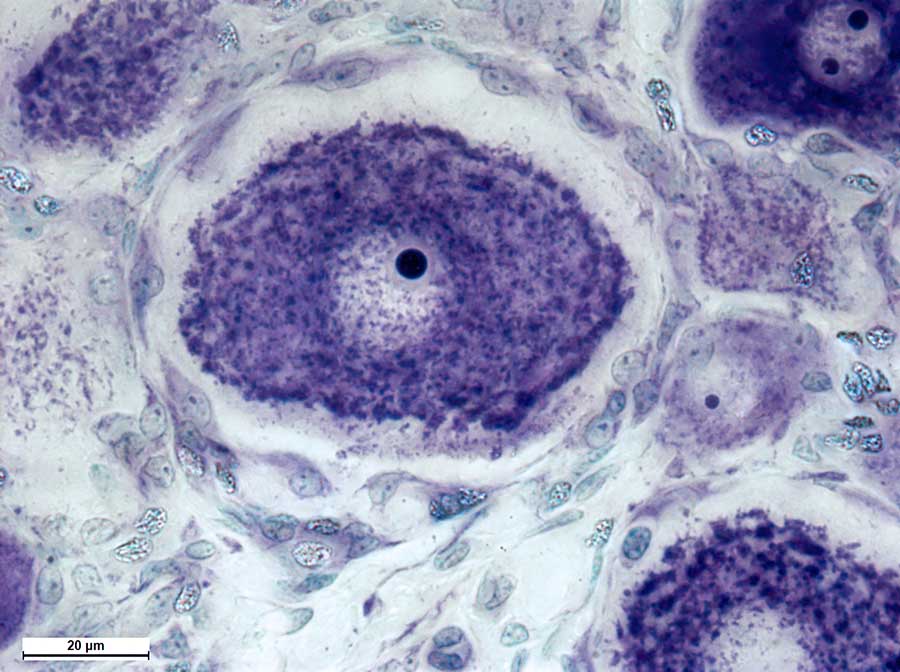
Neurons of a spinal ganglion of a dog. Nissl staining. Photo by O.V. Zaitseva and A.N. Shumeev.
|
||

Multipolar neuron of a peripheral nerve plexus of a chicken. Supravital methylene blue staining. Photo by A.N. Shumeev and O.V. Zaitseva.
|
||
© Laboratory of Evolutionary Morphology (Zoological Institute RAS), IDB RAS, SPBU |
||