
PreparationCross section through a telencephalic hemisphere of the Siberian sturgeon Acipenser baerii Brandt, 1869 (Chordata, Osteichthyes, Acipenseriformes) |
In
|
|
|
Collection SPSU-ODK-PIS-3-1 |
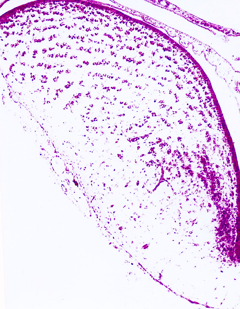
Species: Acipenser baerii Brandt, 1869 Pisces Preparation Type: paraffin sections Prepared: D.K. Obukhov Description: Frontal (transverse) paraffin section through a telencephalic hemisphere of the Siberian sturgeon Acipenser baerii Brandt, 1869 stained with cresyl violet by the Nissl method. Method: Nissl staining Method Description: Frontal paraffin sections stained for Nissl. The staining protocol is described in detail in the handbooks of microscopy methods: Institution: Saint Petersburg State University |
|
|
|
|
|
Series
|
|
|
|
Preparation SPSU-ODK-PIS-3-1 |
Description: Frontal (transversal) paraffin section through a telencephalic hemisphere of the Siberian sturgeon Acipenser baerii Brandt, 1869 stained with cresyl violet by the Nissl method. The section shows the overall cytoarchitecture of an everted-type hemisphere. The section also clearly shows that fishes have only the median ventricle. The neurons are clustered near the surface of the brain and as they migrate deeper into the brain they form a layered structure in the dorsal regions. In the ventral (subpallial) areas, the neurons are even more concentrated near the median ventricle. This type of brain development complicates the comparison with the brains of other vertebrates and suggests that the CNS of this group of fishes (ray-finned fishes) has evolved along its own distinct path. Organs: сentral nervous system, cerebral brain or its part Methods: Neurohistology - Nissl staining |
|