
ImageA group of densely-branched, spiny neurons in the dorsal area of a hemisphere |
||
|
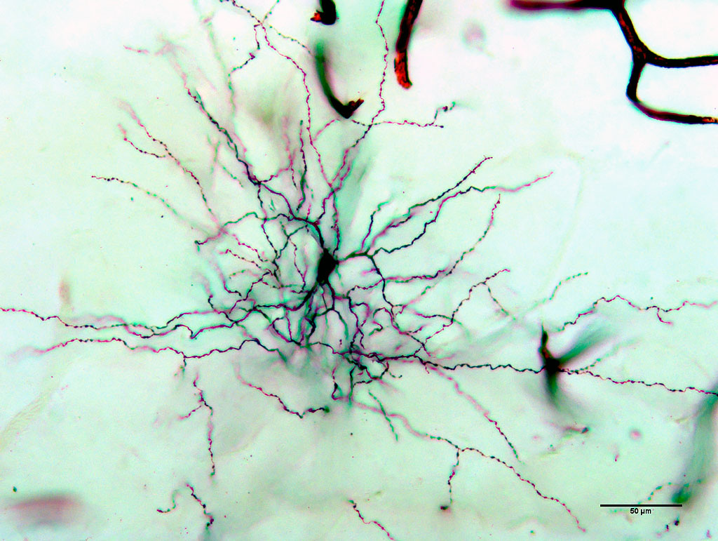
← Click to enlarge image Image Type: Extended focus Projection of optical slices: 1-3 Projections Plane: xyz Color Channels:
Black — silver impregnation License: Open with author and institution Comments: none |
|
Image
|
Frontal (transverse) celloidin section through a telencephalic hemisphere of the common chaffinch Fringilla coelebs L. impregnated with silver salts by the Golgi method. Dorsal region of the pallium (HD – hyperstriatum dorsocellulare). The section shows a separate short-axon, spineless neuron. This is one of the most highly differentiated neurons in the CNS structures of vertebrates, which belongs to a group of highly specialized idiodendritic cells (by the classification of E. Ramon-Molinera). These neurons are usually found in the pallial regions of the telencephalon in Passeriformes (especially in Corvidae) and mammals (in Carnivora and primates). The stack (three images) clearly shows numerous branching axonal collaterals, the soma of the neuron and dendrites (without spines). These cells are interneurons controling the local connections between adjacent neurons in the neuronal complexes (modules). |
|
Annotated
|

|
|