
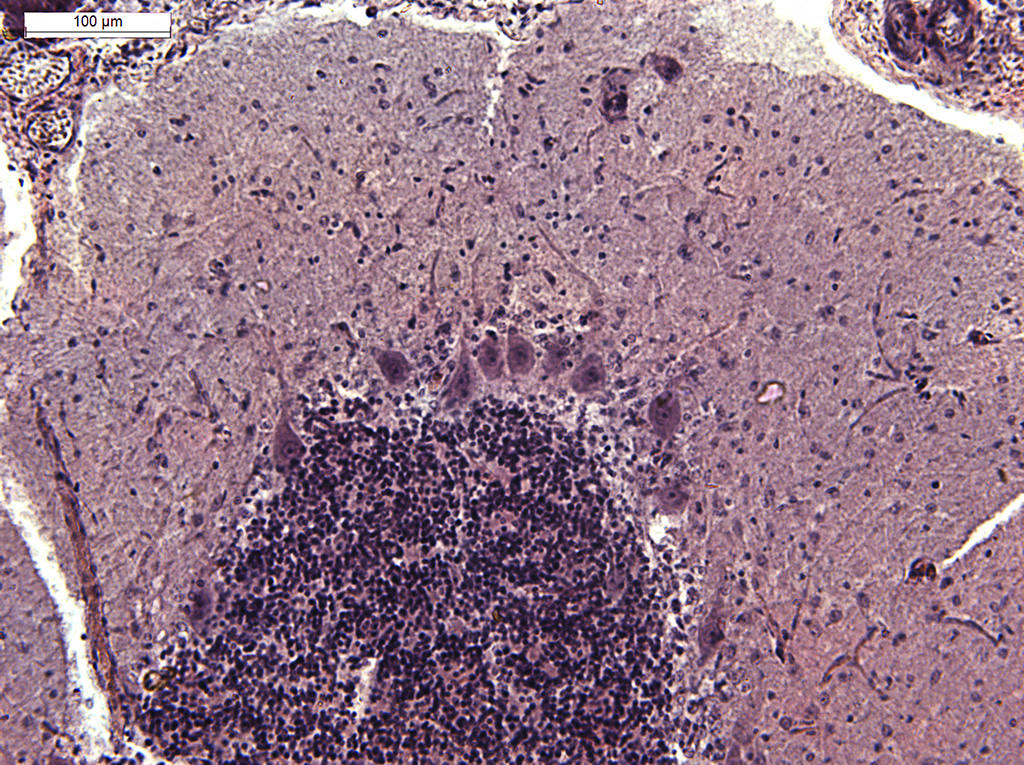
ImageA fragment of a gyrus of the cerebellar cortex of a dog. Hematoxylin-eosin staining. |
||
|
← Click to enlarge image Image Type: Single photo Projections Plane: xyz Color Channels:
Violet — hematoxylin License: Open with author and institution Comments: none |
|
Image
|
General structure of a gyrus in the cerebellar cortex of a dog. The cellular composition of the layers of the gray matter is revealed. |
|
Annotated
|

|
|
© Laboratory of Evolutionary Morphology (Zoological Institute RAS), IDB RAS, SPBU