
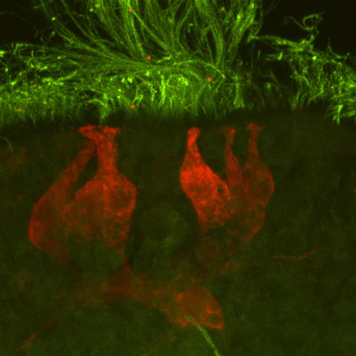
ImageApical organ of a chiton larva Ischnochiton hakodadensis (Mollusca: Polyplacophora) |
||
|
← Click to enlarge image Image Type: Maximum intensity projection Projection of optical slices: 1-80 Projections Plane: xyz Color Channels:
Green — serotonin License: Open with author and institution Comments: none |
|
Image
|
5HT immunostaining of apical organ sensory cells. Cilia are stained with alpha-tubuline. Cerebral commissure is visible underneath the basal parts of the sensory cells. |
|
Annotated
|

|
|
© Laboratory of Evolutionary Morphology (Zoological Institute RAS), IDB RAS, SPBU