
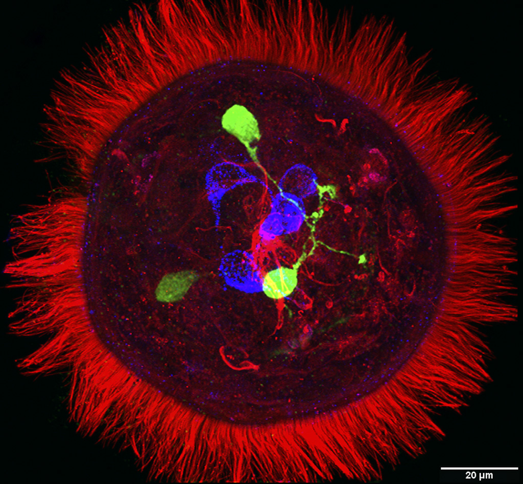
ImageApical organ of a Platynereis dumerilii (Polychaeta, Nereididae) trochophore larva. |
||
|
← Click to enlarge image Image Type: Maximum intensity projection Projections Plane: xyz Color Channels:
Green — serotonin License: Open with author and institution Comments: none |
|
Image
|
Apical organ neurons are located in the center of the episphere. Prototroch cilia are visible along the body edge. |
|
Annotated
|

|
|
© Laboratory of Evolutionary Morphology (Zoological Institute RAS), IDB RAS, SPBU