
A separate sparely-branched, spiny neuron in the central area of a hemisphere of a Japanese quail Annotated Image |
||
|
← Click to enlarge image Authors: D.K. Obukhov Color Channels:
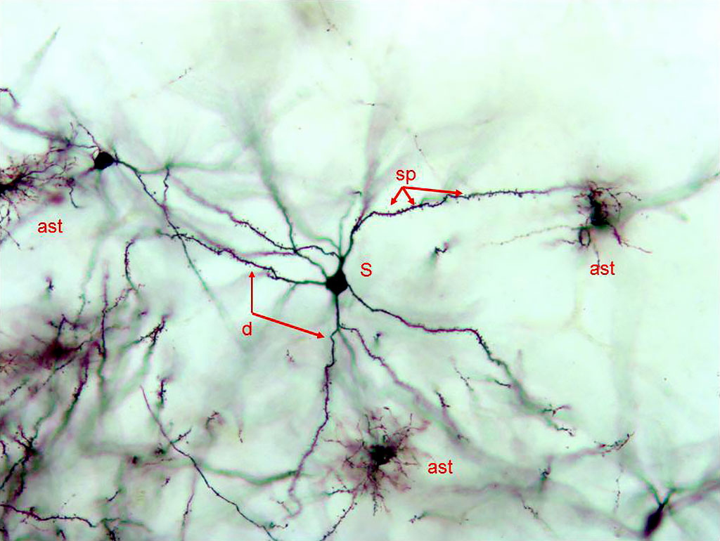
Black — silver impregnation Notation: d – dendrite, |
|
Image
|
Frontal (transverse) celloidin section through a telencephalic hemisphere of a Japanese quail (Galliformes) impregnated with silver salts by the Golgi method (cell bodies and neurites are stained black). Central area of the hemisphere. Silver stained a single sparsely-branched, spiny neuron with numerous spines on dendritic branches (d). According to the classification of E. Ramon-Moliner, these neurons are classified as Class I allodendritic radial neurons. The section also shows capillaries and small blood vessel (bv) and isolated glial cells (presumably the astrocytes - ast). |
|