
Optical
|
||
|
Preparation Storage |
Species: Canis lupus familiaris Linnaeus, 1758 Mammalia Description: With the help of a neurofibril revealing method, bodies and processes of neurons were stained. Large multipolar motoneurons are visible in the gray matter. Comments: Microscope objective 20x Organs: dorsal (spinal) cord or ventral cord, сentral nervous system, neuron(s) Methods: Neurohistology - Cajal's method of silver impregnation of nervous elements Publications: none Microscope: Leica DM4000 B with Leica DFC300 FX |
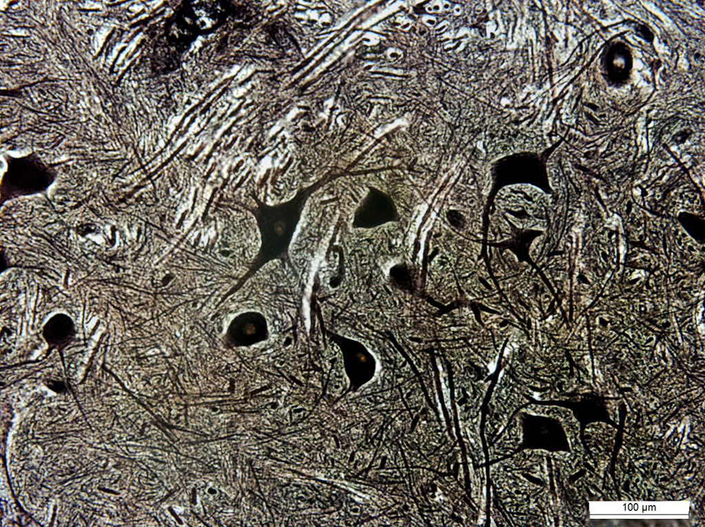
Photo: O.V. Zaitseva, A.N. Shumeev |
All
|
|
|
© Laboratory of Evolutionary Morphology (Zoological Institute RAS), IDB RAS, SPBU