| Craseomys rufocanus (Sundevall, 1846) [n = 1] | [Back to Repository] | |
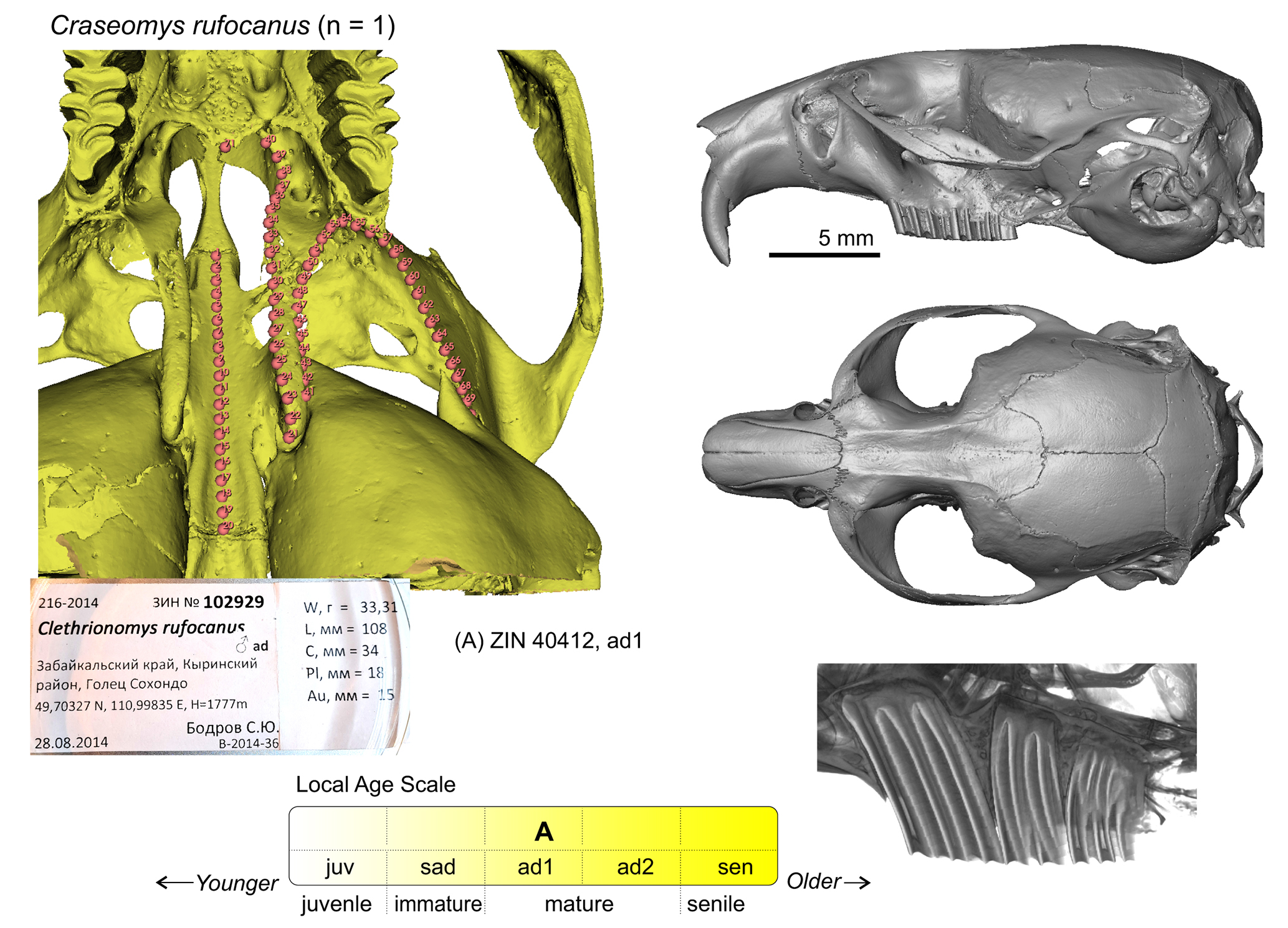
Figure S11. Screenshot of the PSP complex in subposterior view of specimen ZIN 102929 of Craseomys rufocanus with landmark positions (left). Skull in lateral (top, right) and dorsal (bottom, right) view. The lower image shows lateral radiograph of molar roots (root-bearing species only). Scale bar is 5 mm. |
The models licensed under CC-BY 4.0 [Reference: Voyta & Melnikov (2024), DOI: 10.1101/2024.09.04.611334]. Segmentation obtained with Avizo 2019.1 (FEI SAS, Thermo Fisher Sci.) This study was funded by Project No. 19-74-20110 of the Russian Scientific Foundation. PLY files and VTK files can be opened using MorphoDig; VTK files can be combined with Mrk.json files using 3D Slicer.
|
|
| © 2024 zin.ru All rights reserved. Design by Leonid L. Voyta. |